The RBC count method comes under haemocytometry, which quantitatively measures the number of RBCs in a blood sample. Haemocytometer or Neubauer’s chamber slide is a manual method to count RBCs.
Nowadays, more accurate or automated devices have been developed like electrometric and photometric counters that can count the cell constituents of the blood sample.
It is not possible to directly count the RBCs in a blood sample. Thus, it is necessary to dilute the blood sample or blood specimen using one of the RBC diluting fluids (hayem’s or formalin citrate diluting fluid).
In this context, we will discuss the requirements, preparations and procedure of the RBC count through Neubauer’s chamber. You will also get to know the formula for calculating the number of RBCs.
Content: RBC Count Method
What is RBC?
RBCs stand for red blood cells. It is also called erythrocytes, which appears red-coloured due to the coloured pigment (haem) and exists as a biconcave disc. RBCs possess a diameter of 7.5 to 8.7 μm and a thickness of 1.7 to 2.2 μm. It lacks a nucleus and has a life span of 120 days.
Requirements of Total RBC Count
Haemocytometer refers to the micro-slide through which the number of erythrocytes or RBCs can be enumerated via two methods, namely microdilution and macrodilution. Following are the requirements of the RBC count method:
Neubauer’s Chamber or Haemocytometer
It is a specialized thick glass slide used to count the eukaryotic cell suspension. Haemocytometer has a size of 30 X 70 X 4 mm. Its central portion is ruled, where the cell counting is performed. The counting grid has a size of 3 mm X 3 mm. One can estimate the number of red blood cells using a haemocytometer after diluting the blood sample with RBC diluent.
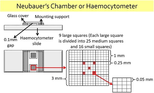
- Nine large squares have an overall width of 3 mm.
- Twenty-five medium squares in each large square have a width of 0.25 mm.
- Sixteen small squares in each medium square have a width of 0.05 mm.
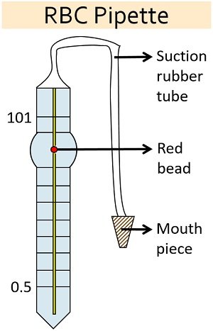
RBC Pipette
It is commonly used to dilute the blood sample with the RBC diluting fluid. It gives a dilution of 1:100 and 1:200. The reading starts from 0.5 to the endpoint of 101. There is a red bead within the RBC pipette, which mixes the RBC specimen with the diluting fluid.
There is a mouthpiece attached towards the end of the suction rubber tube. Through the mouthpiece, the blood is sucked upto a point 0.5 and diluting fluid upto the endpoint 101. The diagram below provides a well-labelled presentation of an RBC pipette.
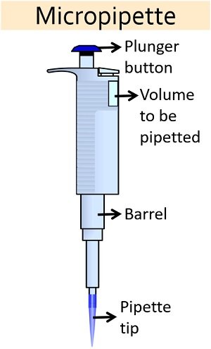
Micropipette
It is a device invented by Dr Heinrich Schnitger. The micropipette is commonly employed in practical or research labs to aspirate or dispense liquid of the desired volume. The size of the micropipette differs.
It uses disposable pipette tips to load or dispense the sample of interest. You could see a diagram below that specifies the parts or components of a micropipette.
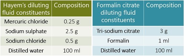
RBC-Diluting Fluid
Hayem’s fluid and formalin citrate diluting fluids are generally used to dilute the RBC specimen. Both function as an isotonic solution, which do not cause haemolysis and the RBCs’ crenation. The composition of Hayem’s and formalin citrate diluting fluid is mentioned below.
Glass Cover
It is a square-shaped coverslip, having a width of 20 mm. To count the number of eukaryotic cells, you should keep the glass cover over the central portion or the ruled area of Neubauer’s chamber. There is usually a 0.1 mm gap between the glass cover and the central area of the haemocytometer.
RBC Specimen
Capillary blood or anti-coagulated blood is generally taken. By pricking the tip of a ring finger, you can directly collect the capillary blood, as capillaries serve as the smallest blood vessels nearby the skin surface.
EDTA serves as an anticoagulant or chelating agent, which prevents blood coagulation by binding with the calcium ions. Other anticoagulants like sodium citrate, potassium oxalate etc., can also be used to prepare whole blood samples.
Video: RBC Count Method
Procedure
To count the RBCs, you can perform microdilution and macrodilution quantitative methods by using Neubauer’s chamber.
Microdilution Method to Count RBCs
Sample preparation: It uses an RBC pipette to incorporate the blood specimen with the diluent.
- Take the blood sample upto a point (0.5). Then, wipe the RBC pipette’s tip using blotting paper.
- After that, suck RBC diluting fluid or diluent upto a mark 101.
- Horizontally rotate the RBC pipette by using your palms.
Loading the sample over the haemocytometer slide:
- Take a clean, grease-free haemocytometer slide and cover glass.
- Place the cover glass on top of the haemocytometer’s lined region.
- Before loading the RBC sample into the haemocytometer, discard 1-2 drops.
- Then, you should carry the RBC pipette at an angle (45 degrees) and load a small volume of RBC sample towards the edge of a cover glass.
- Wait for 3-5 minutes in order to settle down the RBCs in the chamber.
- Observe the prepared slide under the microscope to count the number of RBCs manually.
Macrodilution Method to Count RBCs
- It uses a micropipette to mix the blood specimen with the RBC diluting fluid.
- Take 3.98 ml of RBC diluting fluid plus 0.02 ml of blood specimen (capillary or anti-coagulated blood) into a clean, dry test tube.
- Then, thoroughly mix the contents of the test tube.
- After that, the steps involved in loading a sample over the haemocytometer slide will be the same as the microdilution method.
RBC Count under Microscope
First, focus the rulings of the haemocytometer slide using a 10X objective lens. Using coarse and fine adjustment knobs, focus on the five squares of the large central square to count the number of red blood cells under the 40X objective.
A diagram below represents the pattern to count RBCs in all the five medium squares of a large central square. As already discussed, each medium square possesses 16 small squares. We need to manually count the number of RBCs in five medium squares via hand tally.
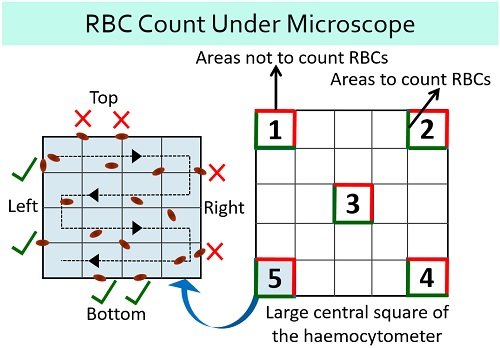
In each square, you need to count the red blood cells located within the square. The red lines in the upper and right corners indicate the areas not to count RBCs, whereas green lines indicate the areas to count the RBCs.
Calculation of RBCs
Total RBCs/µL = Number of RBCs counted X Dilution factor / Area X Depth
- The number of red blood cells (N) =?
- Dilution factor = 1:200 or 200
- A large central square is subdivided into 25 medium squares or sub squares. So, the area will be one sq. mm. The number of RBCs is enumerated in 5 squares out of 25 squares. So, the area of 5 small squares will be 5/25 or 1/5 sq. mm.
- Depth of the sample = 0.1 mm
Total RBCs = N X 200 / 1/5 X 0.1 = N X 200 X 50 = N X 10,000 cells/µL
Suppose, N or number of RBCs in the five squares is 486, then the equation will be represented as:
Total RBCs = 486 X 10,000 = 48, 60, 000 cells/µL
Conclusion
We can conclude that the RBC or erythrocyte count method provides the concentration of RBCs per µL of whole blood. Through a haemocytometer, RBCs are counted manually. RBCs’ normal concentration in children, women and men range between 4.0-5.5 million/mcL, 4.2-5.4 million/mcL and 4.7-6.1 million/mcL, respectively.