Mucor is a mould or a type of fungi, that is found everywhere. There are many species of Mucor (around 50) that are distributed worldwide. It can cause diseases particularly called mucormycosis that might affect the mucous membrane, lungs, eyes, skin etc. Mucor species are fast-growing fungi, which have a highly developed mycelium and branched hyphae. The hyphae in Mucor are generally coenocytic, but septa may appear in the mature hyphae. The cytoplasm of the hypha appears granular.
Classification:
- Kingdom: Mycota
- Division: Zygomycota
- Sub-division: Zygomycotina
- Class: Zygomycetes
- Order: Mucorales
- Family: Mucoraceae
- Genus: Mucor
Habitat: Mucor lives in a habitat like organic soil, a dead decaying matter of fruits, vegetables and plants.
Distribution: Cosmopolitan.
Content: Mucor
Characteristics of Mucor
Some common characteristics of Mucor include:
- Mucor is also called “Black or Bread mould”.
- It belongs to the class of Zygomycetes.
- For most of the Mucor, the mode of nutrition is “Saprophytic” (grows in the dead decaying matter), and for others, it is “Coprophilous” (grows in cow dung or the dung of other herbivorous animals).
- Mucor grows on a variety of substrates like bread, jam, jellies, vegetables etc. The absorption of nutrition is through the mycelial surface or hyphae.
- The vegetative body of Mucor is “Eucarpic” because in this the only thallus differentiates into the reproductive structure.
- The major reserve food material is in the form of glycogen and oil droplets.
- The cell of Mucor is composed of mainly cellulose and chitin.
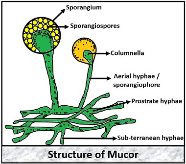
Structure of Mucor
Morphological Features
- Mycelium: The mycelium of Mucor is highly branched, and it forms a fine network of hyphae. A mycelium is simply a cluster of hyphae.
- Hyphae: These are the thread-like and very thin structures that form a “Mycelial network”. Hyphae of Mucor is filamentous, aseptate or coenocytic. In Mucor, the hyphae are of three types:
-
- Sub-terranean hyphae are the type which is highly branched, more penetrating and is present horizontally to the substratum.
- Prostrate hyphae are the type which is also present horizontally between or under the substratum. These two hyphae, i.e. sub-terranean and prostrate hyphae, help in absorption of water and nutrition.
- Aerial hyphae are the type, which originates vertically out from the prostrate hyphae.
- Sporangiophore: It is elongated, slightly narrow in shape.
- Columella: Sporangiophore swells up to form a dome-like structure called “Columella” which can vary in both shape and size.
- Sporangium: It is the round and thick outer covering which carries numerous spores inside it. It can be globose to spherical.
- Spores: These are the reproductive structures forms within the sporangium which are simple, flattened and variable in shape and size.
- Nucleus: Multinucleate nuclei present in Mucor.
Macroscopic Features
- The colony of Mucor shows rapid growth.
- The colour of the colony is usually white to grey and turns to brown when the culture becomes old.
Microscopic Features
- Hypha: Coenocytic and branched
- Spores: Generally black in colour but can vary with different species. The spores can be motile or non-motile and can exist in variable shapes.
Life Cycle of Mucor
It has three modes of reproduction in its lifecycle:
- Vegetative reproduction
- Asexual reproduction
- Sexual reproduction
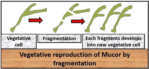
Vegetative Reproduction
It occurs by the fragmentation method, where a vegetative cell breaks into several fragments during some unfavourable conditions. After which, each fragment then develops into a new vegetative body.
Asexual Reproduction
It occurs through the asexual and non-motile spores like:
- Sporangiospores
- chlamydospores
- Oidiospores
Sporangiospores
These are the spores that form within the cell or sporangium and are non-motile. There are following steps involved in the asexual reproduction of Mucor through sporangiospores:
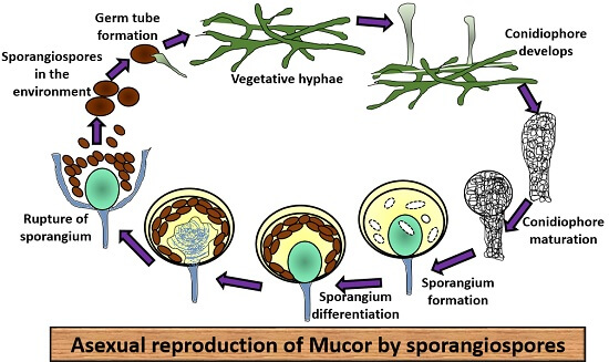
- From the hyphae, first sporangiophores arise singly that are erect in position and unbranched.
- Then, maturation of sporangiophore occurs where the cytoplasm and nuclei push upwards by making the aerial hyphae swollen from the apical end.
- After that, it develops a large round sporangium.
- During the maturation phase, sporangium differentiates into:
- Sporoplasm: It is thick, dense, multinucleate and present inside the sporangial wall.
- Columellaplasm: It is vacuolated and nucleated towards the centre.
- After this, several small vacuoles appear between these differentiated portions. The space between the vacuoles forms cleavage furrows (cavity for cleavage).
- Then, a septum forms to the inner side of the cavity, which further divides into the inner columella and upper sporoplasm. This septum then grows to form a dome shape and later it pushes itself into the sporangium.
- Cleavage occurs in the sporoplasm between the nucleus and the cytoplasm. This division forms a wall around many thin-walled, multinucleate spores called “Sporangiospores”.
- The sporangiospores then releases out of the sporangia after the columnella swells up due to the pressure exerted on the sporangial wall. As a result, cell lysis occurs.
- The spores remain dormant for some time, and when they obtain suitable substratum, they germinate to a new vegetative body through the germ tube.
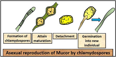
Chlamydospores
A hard wall covers these spores, and it develops inside the vegetative cell during unfavourable conditions. In unfavourable conditions, mycelium becomes septate by the accumulation of nuclei and cytoplasm in a certain portion and becomes surrounded by a thick wall called chlamydospores. This spore then detaches from the mycelium and remains dormant. On favourable conditions, they form a germ tube.
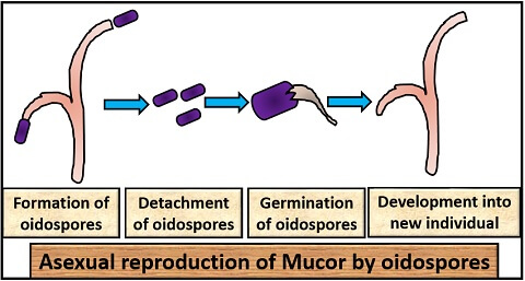
Oidiospores
When a mycelium grows in a substrate (rich in sugar), some small, thin-walled and pearl-like reproductive structures form that detaches out of the vegetative cell as in budding of yeast. Then oidospores remain dormant for some time and on favourable conditions, it forms a germination tube to form a new vegetative body.
Sexual Reproduction
In Mucor, the sexual reproduction occurs by the method that is called as Gametangial conjugation, which involves the following steps:
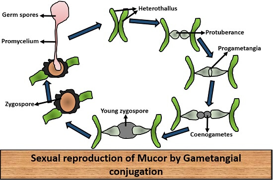
- First, the thallus of two opposite strains, i.e. one is (+), and other is (-), comes in contact with each other.
- When they come in contact, there develops a small outgrowth or protuberance from both of the thalli.
- After that, the outgrowth swells to form “Progametangium”.
- Then septum develops between the progametangium, and the fusion of progametangia occurs that results in the formation of gametes called “Coenogametes”.
- Then gametes of both the strains fuse to form a “Zygote”.
- The zygote then enlarges in size and get surrounded by a thick-walled structure called “Zygospore”.
- Zygospore is dark black in colour, which gets covered by the two layers, namely:
- Outer layer: Also called Exosporium.
- Inner layer: Also called Endosporium.
- The zygospore remains dormant for some time and on favourable conditions, promycelium develops out from the zygospore, forming a new vegetative body.
Through these three reproductive methods, a Mucor completes its reproductive phase, and it can cause some serial infections or diseases that can affect the ecological system and human health.
Thanks. …this is awesome web I got many benefits from this web.
It’s an amazing effort. Very simple and easy concept.
It is an outstanding work. Thanks for helping us.
Thanks a lot for the clear concept and explanation.
Fantastic, thank you so much!
It clears my all concepts. Thank you very much.