Definition: Staining is a method of imparting colour to cells, tissues or microscopic components, so they are highlighted and visualized better under a microscope. There are a variety of staining methods like simple, differential and special staining, which are used for various purposes ranging from the study of microscopic organisms to cellular structures, metabolic processes, cytopathology to name a few.
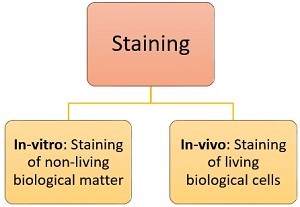
Staining is carried out with the help of a reagent termed as “stain“. This method uses a wide variety of natural and synthetic stains, which is used to add colour to the colourless specimen to be studied. It can be done in two ways, namely in-vitro and in-vivo, that is explained below:
The protocol of staining generally involves three sequential stages:
Smear preparation: This is the primary stage, which involves the mixing of the inoculum with a drop of sterile water and spreading it until a thin film is formed over the glass slide.
Fixation of smear: It is the second stage, which involves drying and heat fixing the thin microbial layer formed on the glass slide.
Staining of the specimen: This is the final stage where the stain is applied onto the dried smear, which imparts colour to the microscopic matter. This procedure is carried out prior to microscopic examination and biochemical tests.
In this article, we will discuss different types of stains, their chemical nature, mode of action and various procedures of staining technique.
Content: Staining
What is Stain?
Stains are chemical reagents or dye that imparts colour to cells and tissue sections of the biological specimens and aids in its visualization under a microscope. Stains work by increasing the contrast between different cellular components, thereby highlighting specific cell structures. Stains can be classified into the following types, depending upon its chemical nature and the type of staining methods.
Based on chemical nature: There are three kinds of stain, acidic, basic and neutral, depending upon the chemical nature of the stain.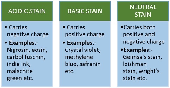
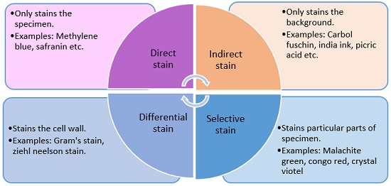
Based on the staining method: There are four kinds of stain, viz. direct, indirect, differential and selective stains.
Purpose
- Enables us to see the organism better: Microorganisms are very minute creatures as well as appear transparent, so staining makes the specimen 9easy to identify.
- Helps to differentiate organisms: Staining helps in distinguishing between the two different groups of organisms, depending upon the colour retaining ability of the cells (some microbes retain the colour of stain, while some don’t).
- To identify a particular structure: For further study of microorganisms, it is also important to study the various internal and external structure of organism like flagella, capsule, nucleus, spores etc.
Mechanism
Stains are organic compound composed of a benzene ring, a chromophore group and an auxochrome group.
Now benzene is a colourless solvent, and the chromophore group is a molecule that imparts colour to the benzene. As a result, the compound formed is called a ‘chromogen’ and it was put forth by O. N. Witt in 1876. Now, this chromogen is not a stain in itself, it is just a coloured compound.
The second part of the stain, the auxochrome, is a chemical group that ionizes the chromogen i.e. it imparts a positive or negative charge to the chromogen group. As a result, the auxochrome enables the ionized chromogen to bind to cells or tissue fibres of opposite charge and thereby colour it.
Types of Staining
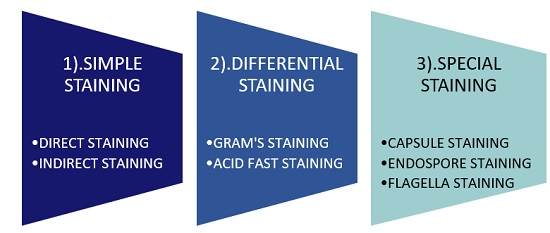
Simple Staining
It determines the cell shape, size and arrangement of the microorganisms. It is a very quick or simple method to perform and it makes the use of a single stain only. These are of two types, namely direct and indirect staining.
Characteristic Differences Between Direct and Indirect Staining:
| Characteristics | Direct staining | Indirect staining |
|---|---|---|
| Stain used | Basic stain | Acidic stain |
| Charge of stain | Positive | Negative |
| Examples | Methylene blue, crystal violet, carbol fuschin | Nigrosine, india ink, congo red |
| Outcome | Stains the specimen | Stains the background |
| General view after staining | 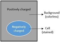 | 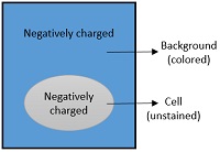 |
| Principle for discoloration | Because of the positively charged stain, it gets attracted towards the negatively charged cell, hence it get fixed to the cell that retain the color of stain results in colorless background with colored cell. | Because of the negatively charged stain, it gets repelled by the negatively charged cell, hence it does not fixed to the cell, results in colorless cell with colored background. |
Differential Staining
It differentiates between the physical and chemical properties of two different groups of an organism, depending on the cell-wall characteristics. It makes the use of multiple or more than one stains. It can be categorised into two types that are given below:
It provides an important tool to differentiate the two major groups of bacteria, i.e. gram-positive and gram-negative. Dr Hans Christian Joachim Gram introduced this method in 1884. It is carried out by the use of differential stain known as Gram’s stain.
Procedure:
| Gram staining | Protocol | Gram positive bacteria | Gram negative bacteria |
|---|---|---|---|
| Primary staining | Heat fixed smear is flooded by crystal violet and allowed to stand for 1min. | ||
| Mordanting | After washing, iodine is then flooded and allowed to stand for 1min. | ||
| Decolourization | After washing, alcohol is added that is washed immediately | ||
| Counter staining | At last, safranin is flooded over the smear and allowed to stand for 30sec, then washed by water. | ||
| Observation | After air drying, place one drop of oil immersion over the smear and adjust the microscope to identify the specimen, whether it is gram negative or gram positive. | ||
| Appear purple in colour because of teichoic acid that resist the primary stain. | Appear pink in color due to lack of teichoic acid,alcohol creates pore in the cell which decolourizes the primary stain |
It differentiates species of mycobacterium from the other groups of bacteria. Paul Ehrlich first developed it in 1882. And later, this technique was modified by a scientist named Ziehl Neelson.
Procedure
| Acid fast staining | Protocol | Acid fast bacteria | Non acid fast bacteria |
|---|---|---|---|
| Primary staining | Heat fixed smear is flooded with carbol fuschin and allowed to stand for 1 min. |  |  |
| Decolourization | After washing, acid alcohol is added. | |  |
| Counter staining | At last, methylene blue is flooded over the smear and allowed to stand for 30 sec, then wash it with water | |  |
| Observation | After air drying, place one drop of oil immersion over the smear and adjust the microscope to identify the specimen, whether specimen is acid fast or not. | | |
| Appears red in colour due to presence of mycolic acid that resist the color of primary stain and does not decolourize. | Appears blue in colour, as they lack mycolic acid, alcohol creates pore in the cell that decolourizes the primary stain. |
Special Staining
It helps in the identification of particular internal and external structural components of the specimen. It includes capsule, endospore and flagella staining.
It differentiates the capsule from the rest of the cell body. This is carried out by the use of both positive and negative dyes.
Capsule: It can define as the polysaccharide envelope, which surrounds the cell wall. Capsule performs many functions like cell protection against desiccation, phagocytic actions and also helps in cell attachment to the host. A capsule is responsible for the pathogenicity or virulence of an organism. It can be seen in the cells of the gram-positive and gram-negative bacteria.
Procedure
| Capsule staining | Protocol | Diagram |
|---|---|---|
| Primary staining | Drop of India ink is placed on a clean slide. | |
| Smearing | Inoculum is then smeared in a dye. | 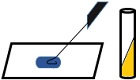 |
| Dragging | Use another slide to drag the mixture into thin film, and then air dried. |  |
| Secondary staining | Crystal violet is flooded over the thin film, and then air dried. | |
| Observation | Examine the cells whether they are encapsulated or not. |  |
| Interpretation of result Positive: Zone formation occurs against dark background Negative: Zone formation does not occur |
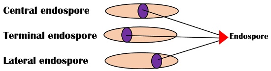
It differentiates the endospore from the vegetative cell and makes the use of both acidic and basic stains.
Endospore: A term itself defines its meaning, in which endo stands for inside and spore stands for a reproductive structure. Therefore, endospores are the reproductive structures inherent to the cell. It acts like a dormant spore, which can resist harsh physical and chemical conditions. Endospores are commonly found in gram-positive bacteria. According to their position, they are of three types as given below:
Procedure
| Endospore staining | Protocol | Diagram |
|---|---|---|
| Primary staining | Malachite green is flooded over the smear | |
| Heat fixing | Then the mixture is heat fixed | |
| Decolourization | Decolourized by water | |
| Counter staining | Safranin is then flooded over the mixture and then air dried | |
| Observation | Examine the slide under the microscope, whether endospore is present or not | |
| Interpretation of result: Positive: If Endospore present, it will appear green in color whereas vegetative cell appears as pink Negative: And if endospore is absent then only vegetative cells will appear pink in color |
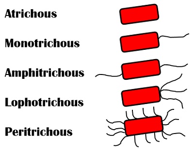
It helps in the identification of the bacterial motility through the presence or absence of flagella. It makes the use of acidic and neutral stain.
Flagella: These are long, thread-like structures, which protrudes outside the cell membrane. Its primary function is to provide motility or locomotion. According to the arrangement, these are of following types:
Atrichous: These are without flagella.
Monotrichous: Single flagellum is present at one end.
Amphitrichous: Single flagellum is present at both the ends.
Lophotrichous: Cluster of flagella are present on one end.
Peritrichous: Flagella are present all over the cell surface.
Procedure
| Flagella staining | Protocol | Diagram |
|---|---|---|
| Primary staining | One drop of leifson’s stain is flooded over the smear | 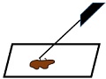 |
| Secondary staining | After that methylene blue is added, and allowed to stand for one minute | |
| Observation | Examine the appearance of flagella to know whether the bacteria is motile or not | 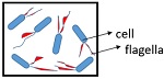 |
| Interpretation of result: Positive: If flagella is present, then it will appear red in color while cell appears blue Negative: And if not present, only cell will appear blue in color |
Examples of Bacteria in different Staining Methods
| Simple staining | Direct stain positive organism: Staphylococcus sp. , E.coli etc | Indirect stain positive organism: Staphylococcus sp. ,Micrococcus luteus etc |
| Differential staining | Gram positive organisms: Streptococcus sp. , Enterococcus sp. , Listeria sp. , Bacillus sp. etc. | Gram negative organisms: Pseudomonas sp. , Salmonella sp. , Klebsiella sp. , Yersinia sp. etc. |
| Acid fast organisms: Mycobacterium sp. | Non acid fast organisms: Enterobacter sp. |
|
| Special staining | Capsule stain positive bacteria: B.anthracis, K.pneumoniae etc. | Capsule stain negative bacteria: Neisseria gonorrhoreae |
| Endospore stain positive bacteria: Clostridium sp. , bacillus sp. etc. | Endospore stain negative bacteria: E.coli , Salmonella sp. etc |
|
| Flagella stain positive organisms: B.subtilis, pseudomonas sp. , E.coli etc | Flagella stain negative organisms: shigella sp. , M.tuberculosis, C.diphtheriae |
Applications
- Staining methods have wide applicability in both biological and biochemical research.
- It is used in staining of metal.
- Used in staining of the wood.
Conclusion
Various staining techniques are used for different purposes like to study bacterial morphology and to examine internal and external cellular components. It can also be used to identify the particular group of bacteria, after which we can further classify the type of specimen, based on their growth behaviour and microscopic characteristics.
I’m grateful for adding another ideal for me pertaining to Staining and stain.
Now that more knowledge concerning that through your website and I will like to know more.
Thanks and God bless u.
This is very helpful to education as a clinical officer
Mam,
First of all, I appreciate the knowledge that you have shared. Best regards!! Thanks for all.
Thank you so much🥰 Such a wonderful deep explanation has been made here. The one who doesn’t know about staining and one who wants to study deep can visit this site it’s really good 👍. Please keep sharing such information, Thank you.
Nice Explanation👍