Colony morphology of bacteria refers to the macroscopic features or cultural characteristics of bacterial masses grown on the nutrient culture media. Bacteria are diverse, and every species have unique culture and growth characteristics.
We can distinguish the colony morphology by looking at the bacterial mass on or in the nutrient base. In microbiology, a colony represents a visible mass of microbial cells.
A single bacterial colony indicates a group of bacterial cells or a bacterial mass. This group of bacterial cells over a nutrient base is what we call a bacterial colony.
In a colony, all the bacterial cells originate from a single mother cell and look identical to each other. By looking into the colony morphology, we can interpret the identity of bacteria.
Different bacteria produce different colonies depending upon shape, size, margin and other features. This post defines the colony morphology and its characteristics. Also, it describes the criteria for recording the macroscopic features of the culture plate.
Content: Colony Morphology of Bacteria
- What is Colony Morphology?
- Colony Characteristics of Bacteria
- How to Observe the Colony Morphology?
- Conclusion
What is Colony Morphology?
Colony morphology is the distinct characteristics of the microbial mass formed on the nutrient base. The distinctive features of a microbial colony include shape, elevation, colour, margin etc.
Example: Staphylococcus aureus in Tryptic soy agar media typically forms circular, convex, golden-yellow colonies with entire margins.
Bacterial Colony
It refers to a bacterial mass derived from a single mother cell. A single mother cell reproduces to give a cluster of genetically identical bacterial cells.
By looking into the bacterial colony morphology, we can identify the type of bacteria. The best way to identify the colony morphology is through microscopic observation.
But before microscopic observation, we can observe the colony characteristics of the bacteria on the agar plate through our naked eyes.
Colony Characteristics of Bacteria
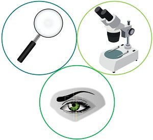
We can view the colony’s morphological characteristics through the following means:
- Naked eye
- Magnifying lens
- Dissecting microscope
There are seven basic characteristics to identify bacterial colony morphology. We can describe the morphology of a bacterial colony by looking into the aspects like:
- Shape
- Elevation
- Margin
- Size
- Colour
- Opacity
- Texture
The above features can be grouped into two categories (form and appearance) that are as follows:
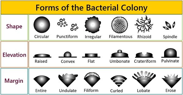
Forms of Bacterial Colonies
It includes features like shape, elevation, and margin of the bacterial colony.
Colony Shape
It refers to the outline of the bacterial colonies. The shape of a bacterial colony we generally encounter are as follows:
- Circular (round shape)
- Punctiform (pin-point or dot-like)
- Irregular (uneven shape)
- Filamentous (thread-like network)
- Rhizoid (branched shape)
- Spindle (cigar-shaped)
Elevation
It describes the height of the bacterial colony above the agar surface. We have to look at the “side view” of a bacterial mass to see the elevation. The most common elevations are:
- Raised (elevated colony above the agar surface)
- Convex (colony is broader at the centre than the edges)
- Flat (uniformly elevated)
- Umbonate (knob in the centre of the colony)
- Crateriform (saucer or goblet-shaped)
- Pulvinate (cushion-shaped)
Margin
It represents the edge of the bacterial colony. The margin of bacterial colonies can be:
- Entire (smooth edges)
- Undulate (wavy edges)
- Filiform (thread-like edges)
- Curled (spiral edges)
- Lobate (finger-like edges)
- Erose (jagged edges)
Colony Appearance
The appearance of a bacterial colony is the way the colony looks. It includes features like size, colour, surface, texture and opacity.
Colony Size
It refers to the colony’s dimensions developed on a culture plate. We can measure the diameter of a bacterial colony in millimetres by using a ruler.
Observation through the naked eye is another way to describe a bacterial mass. Typical shapes of the bacterial colony are:
- Punctiform (<0.5 mm)
- Small (<1 mm)
- Medium (=1 mm)
- Large (>1 mm)
Punctiform colonies are tiny pinpoint colonies. Bacteria having a diameter larger than 5 mm are motile. We can easily measure the colony dimensions from the plate’s base instead of the lid.
Colony Pigmentation
The colony’s colour or pigmentation distinguishes bacteria producing pigments from others. Recording the colour is the first step. Some bacteria impart a distinct colour to the colony.
Examples:
- Pseudomonas aeruginosa produces green pigment.
- Mycobacterium tuberculosis in L.J medium produces buff-coloured colonies.
- Serratia marcescens produce a red-coloured colony.
Opacity or Optical Properties
- Opaque (you can’t see through it)
- Translucent (almost clear, or you can see through it)
- Transparent (clear)
- Iridescent (changing colours in reflected light)
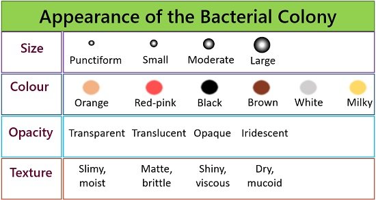
Colony Texture
The texture of the colony is the way a colony’s surface appears and the consistency of the surface. By using an inoculating loop, we can describe the texture of bacterial growth as:
- Dry, mucoid (sticky, mucus-like)
- Slimy, moist (semi-liquid)
- Shiny, viscous (sticks to loop, hard to get off)
- Matte, brittle (dry, breaks apart)
By using a magnifying lens, we can describe the surface of a bacterial colony as:
- Shiny (glistening surface)
- Smooth (lumpless surface)
- Dull (matte surface)
- Veined (lined surface)
- Rough (uneven surface)
- Wrinkled (creased or crumpled surface)
How to Observe the Colony Morphology?
To study the colony morphology of different bacteria, we can expose the nutrient agar plate to varying environments like air, water and soil, etc.
- After overnight incubation, the growth of bacterial colonies in or on the nutrient agar plate occurs. First, look for the separate bacterial colonies and label them as 1, 2, 3 and so on.
- Then prepare an observation table to note down the macroscopic or cultural characteristics of a labelled colony.
- First, record the colour, texture and opacity of the colony you picked.
- After that, notice the desired colony’s size, shape, elevation, and margin.
Besides recording the bacterial colony morphology, it is also necessary to note down the nutrient medium, incubation time and temperature, all of which can affect a colony’s appearance.
Conclusion
Recognizing the colony morphology characteristics is the first step of the identification process. Identifying the different growth morphologies on the nutrient base medium helps us to identify the unknown bacteria. Thus, a systematic assessment of colony morphology is required to indicate that one organism is different from another.