The difference between dicot and monocot leaf is due to the factors like the venation pattern and symmetry.
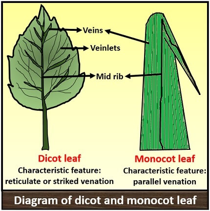
Venation pattern: Monocot leaf shows parallel venation, i.e. in this kind of leaves the veins strike parallel to each other. Dicot leaf shows reticulate venation, i.e. in this the veins of leaf strike towards each other.
Symmetry: Monocot leaf has isobilateral symmetry, i.e. both the leaf surfaces (ventral and dorsal surface) are similar because of an equal number of stomata distribution and undifferentiated mesophyll. Oppositely, the dicot leaf has dorsiventral symmetry, i.e. both the leaf surfaces (ventral and dorsal surface) are different because of a high number of stomata in the lower epidermis and differentiated mesophyll.
Identification features: The dicot and monocot leaf are easily identifiable by observing the presence of stomata and differentiation of mesophyll. A monocot leaf possesses stomata on both the epidermis layers and they generally have undifferentiated mesophyll. Oppositely, a dicot leaf possesses stomata only in the lower epidermis, and their mesophyll is clearly differentiated into spongy and palisade parenchyma.
Content: Dicot Vs Monocot Leaf
Comparison Chart
| Properties | Dicot leaf | Monocot leaf |
|---|---|---|
| Symmetry | Dorsiventral | Isobilateral |
| Shape | Broad or palmate | Long and slender |
| Colour of upper leaf surface | Dark green | Both the upper and lower surfaces are equally green |
| Colour of lower leaf surface | Light green | |
| Veins | Net or reticulate veins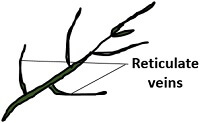 | Parallel veins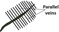 |
| Stomata | Found in lower surface | Equally distributed in both the surfaces |
| Arrangement of stomata | Present randomly | Arranged in parallel rows |
| Guard cells | Kidney shaped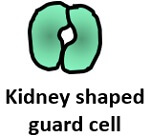 | Dumb-bell shaped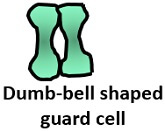 |
| Mesophyll | Differentiated | Undifferentiated |
| Bundle sheath | Single layered | One or more than one layer |
| Colour of bundle sheath | Colourless | Coloured due to abundance of chloroplast |
| Extensions of bundle sheath | Parenchymatous | It is both Parenchymatous and Schlerenchymatous |
| Lateral wall | Sinuous/Curvy | Straight |
| Bulliform/Motor cells | Absent | Present |
| Vascular bundles | Large | Small and large both |
| Arrangement of vascular bundles | Present in rows | Present randomly |
| Intercellular space | Large | Small |
| Silica deposition on epidermal cells | Absent | Present |
| Hypodermis of mid rib | Collenchymatous | Schlerenchymatous |
| Trichome | Absent | Present |
| Examples | Leguminous plants (pea, beans, peanuts etc.), tomato, brinjal, oak leaf etc. | Leaf of grains (Wheat, corn, rice etc.), banana, bamboo etc. |
Definition of Dicot Leaf
The dicot leaves are non-linear, unlike monocot leaves and the vascular bundles in them are irregularly arranged in the net-like veins. These are hypostomatous, i.e. possess stomata on one side (lower epidermis) and have reticulate venation patterns. Dicot leaves comprise the mesophyll differentiated into compactly-arranged palisade and loosely arranged spongy parenchyma cells.
Definition of Monocot Leaf
The monocot leaves are generally linear or oblong and the vascular bundles in them are parallel arranged in the striated veins. These are amphistomatous, i.e. possess stomata on both the sides (upper and lower epidermis) and have striate venation. Here, the mesophyll is undifferentiated.
Diagram
The main characteristic feature to distinguish the dicot and monocot leaf is the type of venation a leaf have. One can easily observe either the veins are striking or parallel by seeing a leaf. Below is the diagram of dicot and monocot leaf, where we can see the venation pattern.
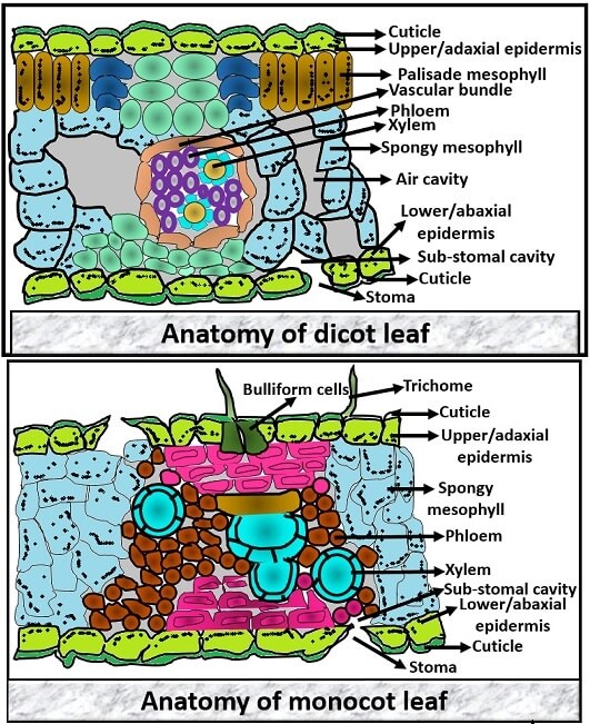
Anatomy of Dicot Vs Monocot Leaf
To know the whole concept of dicot and monocot leaf cell, there are some properties which are as follows:
Symmetry
Dicot leaf shows dorsiventral symmetry where both the dorsal and ventral surface are distinguishable, whereas monocot leaf shows isobilateral symmetry where both the dorsal and ventral surface is similar.
Cuticle
It is the outer waxy envelope that covers the epidermis layer. Dicot leaf has a thin layer of cuticle on both the upper and lower epidermis, whereas monocot leaf has thick cuticle on the upper epidermis and thin on the lower epidermis.
Epidermis
It is a secondary covering that protects the internal cells against damage. It divides into:
- The upper or adaxial epidermis
- Lower or abaxial epidermis
Monocot leaf has the same epidermis layer due to equal distribution of stomata. In contrast, the stomata in dicot leaf are present mostly in the lower epidermis and less or no stomata on the upper epidermis.
Mesophyll
These are the cells that are present below the epidermis. It is the middle layer of a leaf that constitute most of the leaf. It has a high number of chloroplasts that shows photosynthetic activity. In dicot leaf the mesophyll is differentiated into two cells:
- Upper palisade mesophyll: These are elongated cylindrical cells and arranged in the parallel fashion inner to the upper epidermis. Palisade mesophyll contains less or no intercellular space because these lack air cavities.
- Lower spongy mesophyll: These are rounded cells, which are arranged loosely. Spongy mesophyll is present near the lower epidermis. It has large intercellular space because of the presence of air cavities.
Oppositely, undifferentiated mesophyll is present in the monocot leaves. It has only spongy mesophyll, which is spherical in shape and is compactly arranged.
Vascular bundle
It consists of vascular tissues xylem and phloem that plays a significant role in the transportation process. Xylem helps in water transportation which contains vessels, tracheids etc. Phloem contains sieve tubes and companion cells that help in food transportation. The size of the vascular bundle depends upon the size of veins.
In dicot leaf, the vascular bundle is present centrally. The vascular bundle is conjoint, collateral and closed, which is encircled by a single-layered bundle sheath. In monocot leaf, the vascular bundle present parallel in each row. It has collateral and closed vascular bundle that is encased by both parenchymatous and sclerenchymatous bundle sheath.
Stomata and sub stomal cavities
In dicot leaf, stomata and sub stomal cavities are present on the lower epidermis. whereas a monocot leaf possesses stomata and sub stomal cavities on both the upper and lower epidermis.
Bulliform cells
These are the large, colourless, empty cells that attach to the upper epidermis and play a significant function in rolling and unrolling of leaves. In dicot leaf, bulliform cells are absent, whereas present in a monocot leaf.
Key Differences Between Dicot and Monocot Leaf
- The symmetry of dicot leaf is “Dorsiventral”, and in monocot, it is “Isobilateral”.
- A shape of a dorsiventral leaf is broader and comparatively smaller whereas isobilateral leaf is slender and long.
- As the dicot leaf is dorsiventral, the colour of both the upper and lower surfaces will be different in colour. As the monocot is isobilateral, the colour of both the upper and lower surfaces will be green in colour.
- Venation pattern in a dorsiventral leaf is reticulate type, in which the veins are striking towards each other and having net-like veinlets. Oppositely, a monocot leaf has a parallel venation pattern.
- The stomata present randomly and usually on the lower surface of a dorsiventral leaf, whereas, the stomata are present in parallel rows and are uniformly present on both the leaf surfaces of the monocot leaves.
- The guard cells of stomata are kidney-shaped in dicot leaf and dumb-bell shaped in monocot leaf, which is the characteristic feature that differentiates the dorsiventral and isobilateral leaf.
- Another identifying feature is that in dorsiventral leaf, differentiated mesophyll is present (Palisade and Spongy mesophyll) and a monocot leaf has an undifferentiated mesophyll.
- In dorsiventral leaf, a single-layered and colourless bundle sheath is present, while a double-layered and coloured (due to an abundance of chloroplast) bundle sheath is present in the monocot leaves.
- A bulliform or motor cells are present in monocot leaf for the rolling and unrolling action of a leaf, while absent in dorsiventral leaf.
- The vascular bundle is large in dorsiventral leaf, whereas a monocot leaf possesses both large and small vascular bundles.
- Another feature is that in dicot leaf, the intercellular space is more due to the presence of air cavities, whereas the monocot leaves have small intercellular space as they lack air cavities due to the compact arrangement of mesophyll cell.
Conclusion
Therefore, we can conclude that a dicot and monocot leaf differs from each other by the morphological features like venation pattern, symmetry, shape, size, colour and also by the intrinsic feature, i.e. anatomy of the leaf cell.
Do they have different adaptations?
Yes, both dicot and monocot leaves have different adaptations.