The difference between gram-positive and gram-negative bacteria is mainly due to the following factors like:
- Cell wall difference
- Staining difference
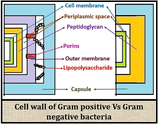
Cell wall difference: Gram-positive bacteria contain high peptidoglycan content (70-80%) and low lipid content (1-4%). It lacks lipopolysaccharide and porins that are present on the outer membrane, and it has less periplasmic space.
Gram-negative bacteria contain low peptidoglycan content (10-20%) and high lipid content (20-30%). The outer membrane is made of lipopolysaccharide and porins, and they have more periplasmic space.
Staining or colour difference: Gram-positive bacteria retain the colour of crystal violet (primary stain) and appear violet in colour due to its thick peptidoglycan cross-linkages.
Gram-negative bacteria lose the colour of crystal violet and appear pink in colour after counterstaining with safranin due to its thin peptidoglycan layer.
Content: Gram-Positive Vs Gram-Negative Bacteria
- Comparison Chart
- Definition
- Cell Wall
- Video
- Colour Difference
- Key Differences
- Similarities
- Conclusion
Comparison Chart
| Properties | Gram positive | Gram negative |
|---|---|---|
| Colour | Appears violet in colour | Appears pink in colour |
| Cell wall thickness | 20-80nm thick | 8-12nm thick |
| Cell wall composition | Low lipid content and high peptidoglycan content (70-80%) | High lipid content (20-30%) and low peptidoglycan content (10-20%) |
| Outer membrane | Absent | Present |
| Teichoic acid | Present | Absent |
| Peptidoglycan layer | Thick | Thin |
| Lipopolysaccharide | Absent | Present |
| Mesosome | More prominent | Less prominent |
| Flagellar structure | 2 rings (S,M) in basal body | 4 rings (L, P, S and M) in basal body |
| Toxins produced | Majority of them produce exotoxins | Majority of them produce endotoxins |
| Porins in outer membrane | Absent | Present |
| Periplasmic space | Comparatively less | More |
| Resistance to physical disruption | High | Low |
| Resistance to cell wall disruption by lysozyme | High | Low |
| Resistance to drying | High | Low |
| Resistance to sodium-azide | High | Low |
| Inhibition by basic dyes | High | Low |
| Susceptibility to penicillin and sulphonamide | High | Low |
| Susceptibility to streptomycin, chloramphenicol and tetracycline | Low | High |
| Susceptibility to anionic detergents | High | Low |
| Pathogenicity | Less bacteria are pathogenic | More bacteria are pathogenic |
| Examples | Bacillus, Streptococcus, Clostridium, Corynebacterium etc. | Enterobacter, Acetobacter, Bortadella etc. |
Definition
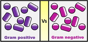
Gram-positive bacteria: These are the type of bacteria, which appear Violet in colour by giving a positive reaction on gram staining, i.e. retain the colour of primary stain (Crystal violet).
Gram-negative bacteria: These are the type of bacteria, which appear Pink in colour by giving a negative reaction on gram staining, i.e. not retain the colour of primary stain and take the colour of counterstain (Safranin).
Cell Wall
The cell wall of gram-positive bacteria is made of sacculus that are the continuous cells, which makes the surface of gram-positive bacteria more even and smooth.
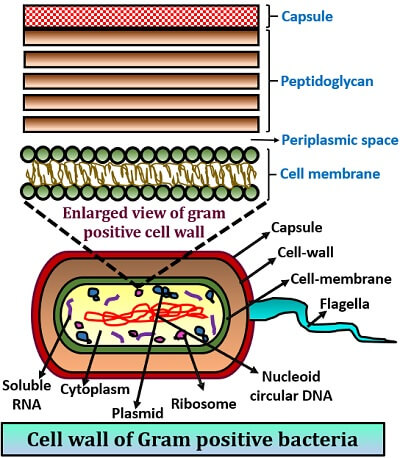
Peptidoglycan constitutes about 70-80% of the cell weight, and lipid content is about 1-4% in the cell wall of the gram-positive bacteria. The cell wall is thick due to a high peptidoglycan content, which is usually multilayered and having dense cross-linkages.
On the peptidoglycan layer of some bacteria, a negatively charged teichoic acid is found. The absence of an outer membrane, porins and lipopolysaccharide in its cell structure is one of the distinguishing features.
Gram-negative bacteria possess a continuous or smooth outer membrane, as it consists of lipopolysaccharide and porins in its structure. It consists of an outer membrane over the peptidoglycan layer, unlike gram-positive bacteria.
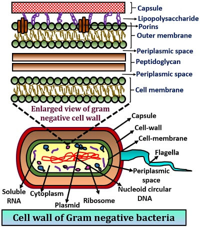
It possesses a high lipid content (about 20-30%) and low murein or polysaccharide content (about 10-20%). The cell wall consists of a thin peptidoglycan layer.
Video: Gram-Positive Vs Gram-Negative Bacteria
Colour Difference
The colour or staining difference between the gram-positive and gram-negative bacteria is due to the retaining ability of the Crystal violet-Iodine complex of the cell wall. This CV-I complex is formed when the Gram’s iodine binds to the crystal violet. The property of the bacterial cell to retain the CV-I complex depends upon the following factors:
- Lipid concentration
- Peptidoglycan thickness
- Presence of magnesium ribonuclease
Colour of Gram-Positive Bacteria
The colour of gram-positive bacteria is violet in colour, as the crystal violet-iodine complex does not come out of the cell during staining. The reasons for the appearance of the violet colour include:
- The gram-positive bacteria contain 1-4% lipid concentration, and by the addition of decolourizing agent (ethanol), the lipid gets dehydrated that the pore size of the cell wall decreases. This is the first reason, by which the CV-I complex cannot come out of the gram-positive bacterial cell.
- The second reason is the presence of a high peptidoglycan content that possesses dense cross-linkages, which strongly holds the CV-I complex.
- Gram-positive bacteria contain magnesium ribonuclease, which covalently binds to the CV-I complex and resists the violet colour.
Colour of Gram-Negative Bacteria
The colour of gram-negative bacteria is pink in colour, as the crystal violet-iodine complex easily comes out of the cell while staining. The reasons for the appearance of the pink colour include:
- The gram-negative bacteria contain a high lipid content (11-20%), and by the addition of decolourizing agent (ethanol), the lipid will dehydrate and the pore size of the cell increases. This is the first reason by which the CV-I complex comes out of the gram-negative bacterial cell.
- The second reason for the elimination of the CV-I complex is due to the thin peptidoglycan layer that comprises only 5-10% of the peptidoglycans cross-linkages.
- It lacks magnesium ribonuclease, by which CV-I complex releases out of the cell.
Key Differences Between Gram-Positive and Gram-Negative Bacteria
- The colour of gram-positive bacteria is violet, whereas the colour of gram-negative bacteria is pink.
- The cell wall of gram-positive bacteria is thick (about 20-80nm), while thin in a gram-negative cell (about 8-12nm).
- The gram-positive bacteria contain 70-80% murein and 1-4% lipid, whereas gram-negative bacteria contain 20-30% lipid content and 10-20% murein.
- Another identifying feature is that the outer membrane, lipopolysaccharide, porins are absent in gram-positive bacteria, whereas present in a gram-negative cell.
- Another main feature is the flagellar structure, where S and M rings are present in gram-positive bacteria. Oppositely, L, P, S and M rings are present in the gram-negative group.
- Majority of the exotoxins are produced by the gram-positive bacteria and endotoxins by the gram-negative bacteria.
- There is less periplasmic space in the gram-positive group of bacteria, whereas more in the gram-negative group.
- Gram-positive bacteria are more resistant to physical dryness, cell disruption, drying and sodium-azide, while the gram-negative bacteria are less resistant.
Similarities
- Both gram-positive and gram-negative bacteria are prokaryotes.
- Membrane-bound organelles are absent in both the groups of bacteria.
- The genome is covalently closed circular DNA in both the types.
- Both the gram-positive and gram-negative groups of bacteria possess the outermost covering, i.e. capsule.
- Peptidoglycan is present in both groups of bacteria.
- The cytoplasm is composed of the lipid bilayer in both of them.
- Both reproduce asexually through Binary fission.
- Both reproduce sexually through transformation, conjugation and transduction.
Conclusion
In this context, we have discussed the differences in the properties like staining and structural properties between the gram-positive and gram-negative bacteria.
This content also gives the idea of the difference in staining properties and the reasons for the appearance of violet and pink colour in of a bacterial cell.
Therefore, we can conclude that both the groups of bacteria are structurally and functionally different due to their cell wall differences, but also have some similarities as they are in the group of bacteria.