Gram staining is used widely and the most popular method in laboratories. It is the type of differential staining, which makes the use of more than one stains to differentiate the bacteria. The gram staining method was first given in 1884 by the Danish scientist and Physician Han’s Christian Gram.
The Gram stain is the differential stain that stains the bacterial cells differently according to the type of cell wall. Gram stain is used to differentiate the bacterial cells by staining the cell wall and distinguish two major groups of bacteria that are gram-positive and gram-negative. Gram-positive bacteria appear violet in colour, and gram-negative bacteria appear pink in colour, as a result of Gram staining.
Content: Gram Staining
Definition of Gram Staining
It is the type of differential staining that is used to differentiate the bacteria majorly into two groups, i.e. gram-positive and gram-negative based on cell-wall difference and by the sequential application of crystal violet, iodine, alcohol and safranin.
What is a Gram Stain?
Gram stain is the differential stain, which makes the use of four reagents that are given below:
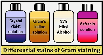
- Crystal violet: It is the basic dye that acts as the “Primary stain”, and it stains all the microorganisms to appear violet in colour.
- Gram’s iodine: It acts as a “Mordant dye” which increases the affinity between the cell and stain by forming a complex with crystal violet known as “CV-I complex”.
- 95% Alcohol: It acts as the “Decolourizing agent” that functions as a lipid solvent, and it dissolves the lipid present in the cell wall. Also, it causes decolourization of the cell by vanishing the CV-I complex from the bacterial cell.
- Safranin: It acts as a “Counterstain” that stains the decolourized bacterial cell by giving it a pink colour.
Video
Principle of Gram Staining
The principle of gram staining relies on the reaction of a bacterial cell with the Gram stain, which finally differentiates the bacteria into gram-positive and gram-negative. First, the Crystal violet dissociates into CV+ and Cl– that further penetrates the bacterial cell and stains the cell violet.
Then, the Iodine (I or I-3) is added, which interacts with the CV+ and forms Crystal Violet-Iodine (CV-I) complex. This CV-I complex forms within the cell wall and the cell cytosol or cytoplasm.
After that, the decolourizing agent, i.e. Ethanol interacts with the lipid content of the bacteria, and this treatment is the important step to differentiate both gram-positive and gram-negative cell.
- In the gram-negative cell, the outer membrane that contains the lipopolysaccharide is dissolved by ethanol. As a result, the peptidoglycan layer gets exposed. The gram-negative cell consists of thin peptidoglycan cross-linkages, by which the CV-I complex is washed out of the cell.
- Oppositely, a gram-positive cell has highly crossed-linked or thick peptidoglycan. This peptidoglycan layer gets dehydrated by the addition of decolourizing agent (Ethanol), which tightens the peptidoglycan layer. This dehydration or tightening of the peptidoglycan layer traps the CV-I complex strongly within the cell.
After decolourization, the gram-negative cells lose the colour of the primary stain and stain pink when treated with a positively charged counterstain, i.e. Safranin. In gram-positive bacteria, the cell retains the colour of the primary stain, i.e. Crystal violet and appears violet in colour.
| Steps in differential staining | Stains used | Gram positive | Gram negative |
|---|---|---|---|
| Primary staining | Crystal violet | Appears violet | Appears violet |
| Mordanting | Gram’s Iodine | Appears violet | Appears violet |
| Decolourization | 95% Alcohol | Appears violet | Appears colourless |
| Counter staining | Safranin | Appears violet | Appears pink |
Process of Gram Staining
The process of gram staining includes the following steps:
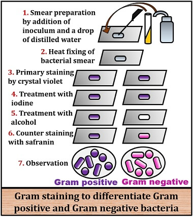
- Take a clean, dry slide and place one drop of distilled water at the centre.
- Prepare bacterial smear by taking little inoculum from the bacterial culture by the help of the inoculating loop.
- Then, mix the inoculum with the water drop to prepare a thin bacterial smear by rotating the inoculating loop in a clockwise and anticlockwise direction.
- After that, the bacterial smear is heat-fixed under the spirit lamp.
- Then, flood the bacterial smear with crystal violet and allow it to stand for 1 minute.
- Then flood the smear with Gram’s iodine.
- After that, add the decolourizing agent, i.e. 95% Ethanol to the smear.
- At last, add counterstain, i.e. Safranin to the smear and allow it to stand for 1 minute.
- Then, wash the slide with water and air dry the slide.
- At last, observe the glass slide under the microscope by adding oil immersion to the stained cells to differentiate gram-positive and negative cells.
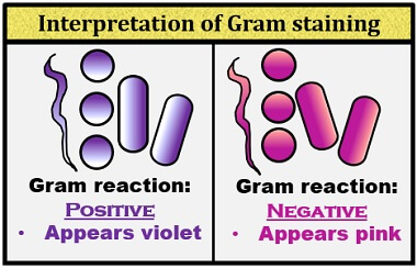
Result Interpretation of Gram Staining
- Gram-Positive: If the bacteria cell gives a positive result for the gram reaction, it will stain Violet.
- Gram-negative: If the bacterial cell gives a negative result for the gram reaction, it will stain Pink.
Significance
Gram staining is the primary step for the initial identification and classification of the bacteria into two groups, i.e. gram-positive and gram-negative. It stains the bacterial cell wall differently, by giving violet colour to the gram-positive bacteria and pink colour to the negative bacteria. Therefore, it adds colour to the cell wall of bacteria, which increases the visibility under the light microscope.
Besides, gram staining also helps in the study of the morphology and arrangement of the bacteria. It also helps us to know about the chemical and physical properties of the gram-positive and gram-negative bacteria, based on the cell wall difference.
Why do we need fresh culture to perform gram staining???