Mu phage is also called temperate or transposable phage because it causes transposition of the genes into the host cell, at the time of multiplication. It mainly attacks the members or species of enterobacteria family, and that’s why also known as “Enterobacterial phage Mu”. The transposition of Mu-phage occurs by either of the two mechanisms:
- Replicative transposition
- Non-replicative transposition
Host range: E.coli, Serratia marcescens, Citrobacter freundii, Erwinia carotovora etc.
Classification:
- Group: Group-I (ds-DNA)
- Order: Caudovirales
- Family: Myoviridae
- Genus: Mulikevirus
Content: Mu Phage
Meaning
It is a type of a bacteriophage (the virus that invades the host bacterial cell), which shows the properties of both viruses and transposons.
History
Several discoveries have led to the study and discovery of Mu-phage.
| Year of discovery | Discoverer | Discovery |
|---|---|---|
| 1950 | Larry Taylor | Discovered the Mu-phage. |
| 1972-75 | Ahmad Bukhari | Demonstrated the insertion of Mu randomly throughout the bacterial genome. Introduced the host gene by the excision of Mu. |
| 1979 | Jim Shapiro | Introduced transposition model as “Shapiro- intermediate”. |
| 1983 | Kiyoshi Shapiro | Demonstrated in vitro transposition by using mini Mu-plasmids. |
| 1994-2012 | Montano | Created crystal structure of the Mu-bacteriophage transposome. |
Structure of Mu Phage
The basic structure of the bacteriophage possesses head, neck and tail. Therefore, the structure of Mu phage is also divided into three parts, i.e. head, neck and tail. To know more about the structure of Mu phage, let us look at the picture given below.
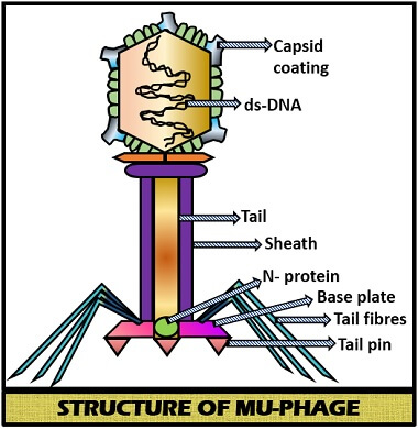
Head: It is the terminal part of the Mu phage, which carries the viral genome and a protein later (capsid) encases the hexagonal head.
- Symmetry: The head of Mu phage possesses icosahedral symmetry.
- Shape: The shape is like spheroid.
- Diameter: The diameter is 54 nm.
- Capsid: It is the layer which surrounds the hexagonal head. The capsid of Mu phage is composed of about 152 smaller protein subunits, known as capsomeres.
Neck: It is the middle portion of the Mu phage, which acts as the joining element of the two components, i.e. head and tail.
Tail: It is long, thick and contractile because of the presence of crossbands.
- Size: The size of the tail is 183 X 16-20 nm.
- Sheath: It is the covering of the contractile tail, which is composed of stacked rings. At the time of contraction, the sheath becomes shorter and thicker. And the length of the sheath during contraction is up to 60-90 nm.
- Tail fibres: These are thin and thread-like structures, which are attached to the large base plate. There are six long terminal fibres.
Genetic Map
Mu phage consists of a ds-DNA that is linear and non-segmented. Its genome consists of 37,611 base pairs. The guanine and cytosine content is 35%. The genome is non-permuted, consists of some unusual base pairs and terminally redundant sequences. Mu phage genome encodes 55 genes, which perform different functions in its life cycle. The genome size is 40 Kb in length.
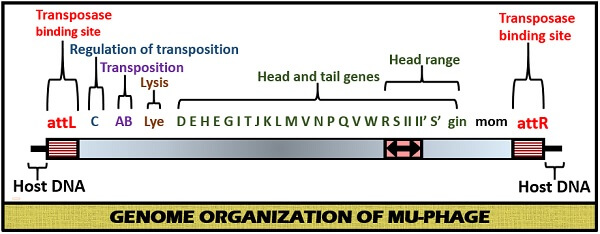
The functions of different genes are as follows:
- Mu-phage consists of two transposase binding sites that bind to the host DNA and represented as attL and attR. These two ends sometimes called as MuL and MuR.
- A gene: It encodes all transposition events.
- B gene: It encodes the all replicative transposition.
- C gene: It represses the expression of the transposase gene.
- Head and tail genes: These are the structural genes that help in reconstruction or biosynthesis of Mu phage.
- Gin gene: It catalyzes the site-specific inversion reactions.
- Mom gene: It encodes a DNA modification function by converting adenine to acetamide adenine.
- Lye gene: It encodes the lytic enzyme, which causes lysis of the host cell.
Life Cycle of Mu Phage
Its lifecycle can be summarized in the following steps:
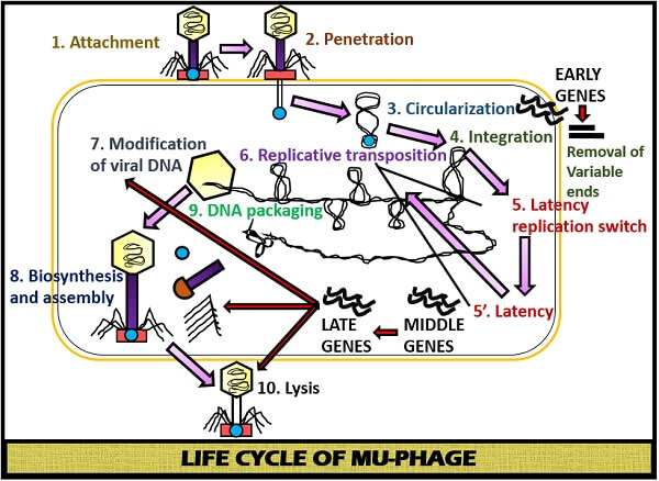
Attachment
Firstly, the tail fibres attach to the receptor site of the host cell surface. By the binding of the tail fibre, there is the conformational change in the base plate of Mu phage. Due to the conformational change in the base plate, the tail’s sheath contracts.
Penetration
By the contraction of the tail’s sheath, the rigid internal material gets into the host cell surface through the cell envelope. The N protein ( non-replicative protein) also gets injected along with the viral genome.
Circularization
The N-protein undergoes circularization once it binds with the viral genome.
Integration
After circularization, early transcription occurs that gives rise to the Repc and Ner repressors and DDE recombinase A (Mu A). These genes help in the integration of the viral genome with the host genome. During this step, the variable ends are cut off from the viral genome.
Early phase
After the non-replicative transposition, the ratio of Repc and Ner repressors decide whether the phage will enter to the lysogenic phage or lytic phage.
- Repc: It represses the early promoter by establishing latency or lysogeny.
- Ner: It represses the expression of Repc by promoting the expression of the early genes for the replication of Mu phage.
Middle phase
After the inactivation of Repc, there is an expression of MuA and MuB genes. MuA is the DDE recombinase-A enzyme, and MuB is the target DNA activator B. MuA performs the transposition of viral genome ends and host DNA. The target DNA activator B helps in the replication of viral host DNA, leading to the formation of the two copies. This type of replication is called replicative transposition. This replication can lead to 100 viral genomes after successive rounds.
Late transcription
This phase carries out the expression of the adenine modification enzyme, which makes the viral DNA resistant to the host restriction enzymes by modifying the adenines in the viral DNA.
Biosynthesis and Assembly
The late gene synthesizes the structural genes of Mu phage, which leads to the biosynthesis of virus particles. Then the virus particles like empty capsid, tail fibres etc. get to assemble.
The packaging of virion
Firstly, the bacterial DNA is first cut on the left of the integrated Mu genome for about 50-150bp. Then, a second cut occurs after the filling of phage head. The packaging of viral DNA also occurs on the right side of the Mu genome. Therefore, at different sites of the bacterial genome, the packaging of the Mu genome will occur.
Cell lysis and release of virion
After packaging, the newly synthesized virions release out of the host cell by the help of lye gene that encodes lytic enzymes (responsible for the cell lysis).