The WBC or leukocyte count method estimates white blood cells per microlitres of your blood. By enumerating the total number or concentration of leukocytes, you could determine the condition of your immune health. The average WBC count is between 4000 to 11000 cells/µL of blood.
An increase in the concentration of leukocytes leads to a medical condition called “Leukocytosis”. Oppositely, a decrease in the concentration of leukocytes eventually leads to “Leukopenia”. Therefore, white blood cells make a significant difference to health, as immunity depends on it.
The total WBCs in the blood smear is called WBC count. It involves counting WBCs manually using a haemocytometer and automated WBC counters using impedance, quantitative buffy coat, and flow cytometry techniques. Here, we will discuss the manual method to count white cells.
The principle of the WBC count method depends upon the dilution of either anticoagulated blood or capillary blood, using WBC diluent. Then the cover glass is mounted over the diluted blood smear in the central ruled area of the Neubauer’s chamber.
Finally, the prepared slide is examined under the light microscope to count the total number of WBCs within the four large corner squares of the haemocytometer. This post describes the meaning and types of WBCs. You will also get to know the requirements, procedure (Thoma pipette method and tube method), criteria to count WBCs under the microscope and the calculation of WBCs.
Content: WBC Count Method
Meaning of WBC
WBCs, leukocytes or white blood cells are the immune cells contributing 1% of your blood. Hematopoietic stem cells of the bone marrow form white blood cells that flow through the bloodstream. Spleen, liver and kidneys regulate the WBC’s production. The blood and lymphatic system carry the leukocytes.
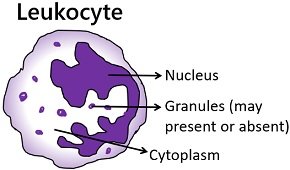
Unlike RBCs, white blood cells possess nuclei and lack haemoglobin. Based on the presence of granules, there are five types of WBCs.
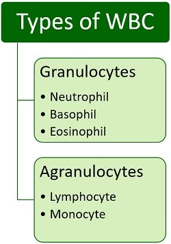
- Neutrophils have a segmented nucleus and contain light pink coloured granules in the cytoplasm. These play a crucial role to phagocytose bacteria and promoting inflammation.
- Basophils have a bilobed nucleus and dark blue or purple coloured granules in the cytoplasm. They mediate allergic responses.
- Eosinophils have a bilobed nucleus and contain dark pink or red coloured granules in the cytoplasm. These are active against parasites and helminths.
- Lymphocytes have a large spherical nucleus with agranulated cytoplasm. These include effector and regulatory cells (like T, B and NK cells) that trigger an immune response.
- Monocytes have a large indented nucleus with agranulated cytoplasm. These play a significant role to form macrophages and other antigen-presenting centres.
Requirements of WBC Count
To manually count the white blood cells, you need the following devices and reagents:
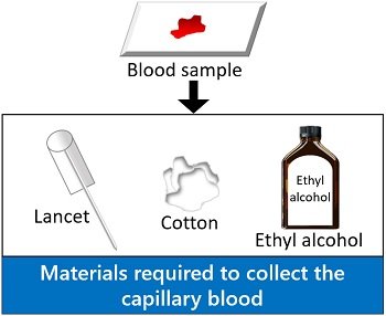
Materials required to collect the capillary blood
Cotton, lancet, ethyl alcohol and glass slide are the materials needed to collect the blood sample. We will discuss the process of collecting blood further in this post.
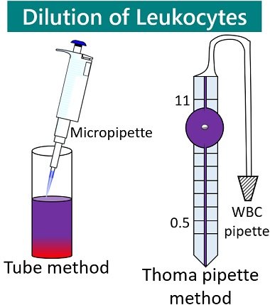
Materials required to dilute blood
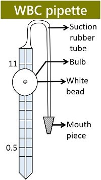
WBC Pipette: It is also called Thoma-pipette. WBC pipette is an apparatus used to prepare the TLC (total leukocyte count) solution. It gives a dilution of 1:20. The stem of the WBC pipette has markings 0.5 to 1.0. A white bead is present in the swelled central portion or bulb of the WBC pipette. The short arm after the bulb is marked with the endpoint 11. There is a rubber tube with a mouthpiece, through which you can suck the blood.
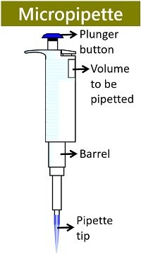
Micropipette: It is required to aspirate or dispense the blood sample and WBC diluent of the desired volume into the sterile test tube. In the bulk dilution method, a micropipette is an apparatus used to dilute white cells with the WBC diluent.
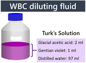
WBC Diluting Fluid: Turk’s solution dilutes the blood specimen, and it has the following components:
- Glacial acetic acid: 2 ml
- Gentian violet: 1 ml
- Distilled water: 97 ml
Gentian acetic acid causes lysis of erythrocytes. Gentian violet stains the WBC’s nuclei. Distilled water is used to dilute the contents of the Turk’s solution.
Haemocytometer
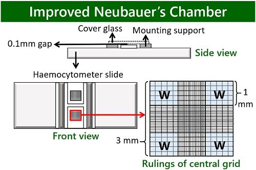
It was pioneered by Louis Charles Malassez to count blood cells. Haemocytometer is a thick microscope slide with three lateral platforms. It possesses four vertical troughs and one horizontal trough. The central platform has an etched grid at the centre.
Haemocytometer has an H-shaped volume chamber. Raised edges of the haemocytometer hold the thick cover glass at the height of 0.1 mm from the ruled grid. The improved Neubauer’s slide has nine large squares in a ruled grid, among which four large squares at the corners are examined to calculate WBCs per microliter of a blood sample.
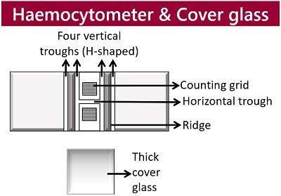
Cover Glass
It is a specialized coverslip that is thicker than ordinary coverslips.
- Thick: 0.4 mm
- Width: 26 mm
- Length: 20 mm
The special cover glass of the haemocytometer slide is placed 1 mm above the blood specimen loaded into the counting grid.
The Procedure of Total Leukocyte Count
The Thoma pipette technique and tube method are commonly used to count the white blood cells manually. Both use the same dilution of the blood specimen. Let us summarize the steps of both methods.
Collect Blood Sample
You can use capillary blood directly by pricking the fingertip. First, rub the tip of your middle finger with cotton saturated with ethyl alcohol. Then, prick the tip of your finger by using a sterile lancet. Put 2-3 drops of your blood over the centre of a sterilized glass slide. The procedure of collecting a blood sample is similar in both the Thoma pipette method and the tube method.
Dilution of Blood Specimen
Thoma pipette method: Suck blood up to the 0.5 mark through the mouthpiece attached to the end of a rubber tube. Afterwards, you need to suck the prepared Turk’s solution (WBC diluting fluid) up to an endpoint 11 in a similar way. Then, horizontally rotate the WBC pipette using your palms in order to mix the contents.
Tube method: Dilution of blood specimen in the tube method is quite different, as it uses a test tube to mix the blood sample with WBC diluent instead of using a WBC pipette. The dilution factor is the same (1:20) as the Thoma pipette method. You need to add 0.02 ml of blood into the sterile test tube using a micropipette. Similarly, dispense 0.38 ml of WBC diluting fluid into a test tube. Then, vigorously shake the test tube to mix the contents.
Sample Loading and Slide Preparation
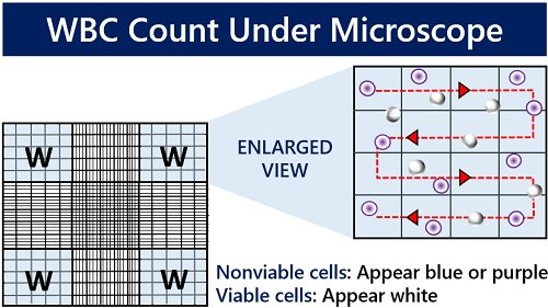
Thoma pipette method: Before loading the sample into the central ruled portion of the haemocytometer slide, you must discard a few drops from the WBC pipette. Place a thick cover glass over the central grid with a space leaving about 1 mm. Then, carefully load the sample from both the edges of a cover glass. Through capillary action, the sample reaches the central ruled area of the haemocytometer. Finally, allow the cells to settle for at least 2 minutes. In the diagram, the areas highlighted in blue are the WBC counting regions that are denoted by ‘W’.
Tube method: Charge the diluted blood specimen to fill the central ruled portion of the haemocytometer using a micropipette. Else, you need to follow the same steps of the Thoma pipette method to prepare the slide for microscopic observation.
How to count WBCs?
First, you need to focus on the rulings of the central grid under the 10X objective. Then, focus on the four large squares at the corners by adjusting coarse and fine adjustment knobs. Then, manually count the leukocytes within four corner squares of the ruled grid via hand tally.
Calculation of WBCs
After counting white cells in four large corner squares, we can put the values in the given formula:

- Numbers of WBCs counted = ‘N’
- Dilution factor= 20
- Depth = 10
- Area= 4 mm2
Hence, WBCs/µL= N X 10 X 20 / 4 = N X 50 cells/µL
Suppose the number of cells in the first and second grid is 160 and 200, respectively. To find out the total number of WBCs, you need to sum up the above two numbers and multiply them by 50. Thus, we could get the total leukocyte count by putting the final value in the equation.
WBCs/µL= 360 X 50 cells/µL = 1800 cells/µL
Conclusion
Therefore, we can conclude that the WBC count method is a form of blood test through which we could estimate the concentration of leukocytes. Thus, TLC is a quantitative measurement of white blood cells manually through a wet mount.
Awesome explanation with very very small details.