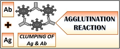
Agglutination reaction is an immunological assay, which results in the specific antigen and antibody reaction in vitro. Agglutination purely refers to the clumping or aggregation of the particles. As a result of agglutination, a cross-linked structure or lattice appears in the form of visible aggregates.
Agglutination reaction is a sensitive, easy and cost-effective method to carry out and does not require technical skills for the operation. The lattice or the antigen and antibody network can be seen by macroscopically and microscopically depending upon the type of agglutination reaction. Here, we will discuss the definition, history, principle, steps, types and application of the agglutination reaction.
Content: Agglutination Reaction
- Definition of Agglutination Reaction
- History
- Principle of Agglutination Reaction
- Steps of Agglutination
- Types of Agglutination Reaction
- Applications
- Conclusion
Definition of Agglutination Reaction
Agglutination reaction is a serological assay, which results in the clumping of reactants (antigens and antibodies) to form a large visible aggregated mass. It involves the mixing of large or particulate antigens with the antiserum containing antibodies over the solid matrices like glass side, microtitre plate or test tubes.
The agglutination between antigen and antibody occurs at the optimal temperature, pH and ionic strength of the solution. The combination of both the reactants, i.e. antigen and antibody result in the formation of antigen-antibody network or lattice as “visible clumps”. The most common example of the agglutination reaction is “Blood typing”.
History
| Year | Discoverer | Discovery |
|---|---|---|
| 1896 | Herbert Edward Durham and Max Von Gruber | Introduced the theory of “Specific- agglutination” |
| 1862-1926 | Fernand Widal | Introduced Widal agglutination for the diagnosis of typhoid fever |
| 1900 | Karl Landsteiner | Introduced blood typing based on the principle of “Agglutination” |
Principle of Agglutination Reaction
The principle of agglutination reaction is based on the “Clumping of antigen and antibody”. Like precipitation reaction, it also involves the binding of antigen and antibody at the equivalence zone, where the concentration of both are at equilibrium. Antibodies possess a y-shape with two Fab sites made of the hypervariable region, which target the specific antigenic determinants or epitopes of an antigen.
The binding of antigen and antibody is similar to the “Lock-key model”. Therefore, we can take a reference by considering the epitopes of an antigen as a key and the Fab sites of an antibody as a lock. Thus, the specific epitope will fit into the cleft of an antibody’s Fab sites.
Steps of Agglutination
Agglutination reaction involves two steps:
- Sensitization: It is the primary stage where binding of antigen and antibody takes place. Temperature, pH, ionic strength and incubation period influence the efficiency of sensitization or binding. Sensitization is not a visible reaction.
- Lattice formation: It is the secondary stage where the antibody and multivalent antigen forms a stable network called “Lattice”. A lattice exists a net-like configuration, which consists of a stable network between sensitized antigen and antibody. It takes much time to occur than the sensitization. Lattice is a visible reaction.
Types of Agglutination Reaction
Active and passive agglutination are the two common agglutination assays, based on the nature of the antigen.
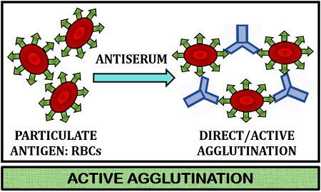
Active Agglutination
It involves direct interaction of antibodies present in the serum with the particulate antigens carrying epitopes of interest. Active agglutination is used to quantify antibodies against the particulate antigens like RBCs, pathogenic microorganisms like bacteria, fungi etc.
In this method, add the particulate antigens into the wells of a microtitre plate. Then, serially dilute the antibodies and pour the suspension into the wells coated with an antigen. By increasing the concentration of antibody, the rate of agglutination will also increase.
- Positive result: Antibodies present in the serum will bind with the particulate antigen and results in the formation of the antigen-antibody mat at the bottom of the well.
- Negative result: It occurs due to an insufficient amount of antibodies to agglutinate the antigens. Therefore, the agglutination reaction will not occur. In this case, the antigens would appear as pellets at the bottom of the well, instead of aggregates.
Example: Slide agglutination test(Blood-grouping), Widal test for the diagnosis of typhoid fever, Coomb’s test to detect anti-Rh antibodies etc.
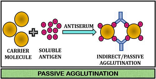
Passive Agglutination
It involves indirect interaction of the antibodies with the soluble antigens via carrier particles. Passive agglutination primarily involves the binding of soluble antigen with the carrier matrices like latex bead, polystyrene etc. In this type, the antigens do not carry epitopes of interest, which have to be agglutinated.
The another type (reverse passive agglutination) uses known antibodies in place of antigen, which attache with the carrier matrix. After the antibody’s attachment with the carrier molecule, the antigens attach to the Fab sites of the antibody to agglutinate it.
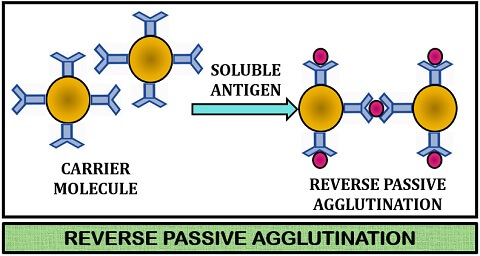
Example: Latex bead agglutination for both antigen and antibody.
Applications
Agglutination test is widely applicable in the field of clinical microbiology, and some of the applications are given below:
- Blood typing of recipient and donor at the time of blood transfusion.
- Helps in the detection of antibody presence and to quantify the amount of antibody present in the patient’s blood.
- Active agglutination helps in serodiagnosis of toxoplasmosis, brucellosis etc.
- Passive agglutination helps in the determination of the Rh-factor.
- It is extensively used in techniques like latex and haemagglutination.
- It helps to detect the type of antigen and quantify the amount of antigen present in the patient’s blood.
Conclusion
Therefore, active and passive are the two main types of agglutination based on an antigen’s nature. Other than active, passive, and reverse types, there is another type called miscellaneous agglutination. The clumping occurs between the antibodies against a particulate antigen of different, but the related organism results in cross agglutination.
Agglutination between the antigens of the biologically related organism and the specific antibodies is called group agglutination. The clumping of particulate antigenic elements with the blood cells like RBCs within the blood vessels causes intravascular agglutination.