The anatomy of the rib cage in the human body contains twelve pairs of ribs, twelve thoracic vertebrae with one sternum and ten costal cartilages. It is a semi-solid bony and cartilaginous structure.
The rib cage is also called the thoracic cage. It makes up the thorax or chest segment of our body, which exists between the neck and abdomen.
The skeleton around the thorax is an osteocartilaginous cage. It is a part of the axial skeleton. The rib cage protects and supports the underlying organs in the thoracic cavity, i.e. lungs and heart.
Thus, it aids in respiratory and circulatory mechanisms. Rib cage generally seems narrow superiorly and broader inferiorly.
Here, one interesting fact is that women have smaller ribcages than men.
This post describes the meaning and anatomy of the rib cage. Also, you will find the parts and function of each component of the thoracic cage.
Content: Anatomy of Rib Cage
Meaning of Rib Cage
It is a part of a rib-vertebral-sternal complex surrounding the thorax of the human body. The thoracic cage includes twelve pairs of ribs. These pairs attach to the corresponding sites of the sternum and the thoracic vertebral column.
- Posterior view: The rib cage appears flattened posteriorly. The posterior T1 to T12 section of the vertebral column forms a pair of twelve ribs.
- Anterior view: It has a dome shape. The first ten ribs move anteriorly to the sternum or breast bone via hyaline costal bones. And the last two ribs remain free.
Rib Cage Anatomy
We will discuss each component and structure of the rib cage in detail in the upcoming sections. But, before proceeding to that, let us take a brief overview of the rib cage anatomy.
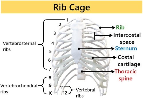
The thoracic spine supports the rib cage posteriorly. A human body has 12 thoracic vertebrae to which the head of 24 ribs articulates with the vertebral body’s demi facets. Whereas, the tubercle of the 24 ribs articulates with the costal facets of the vertebral arch.
Humans have 24 ribs that curve towards the anterior part of the sternum.
- True or vertebrosternal ribs (1 to 7 pairs) directly articulate with the sternum’s specific region.
- In contrast, the thoracic cage has 8 to 10 pairs of false ribs. The 8th to 10th pair of ribs connect with the costal cartilage of the seventh rib.
- The 11th and 12th pairs of ribs are the floating ribs. These ribs have no connection with the costal cartilage and sternum.
Intercostal space is the vacant space between the two adjacent ribs. Nerves, lymph nodes, arteries, veins and muscles occupy the intercostal spaces. The sternal ends of 20 ribs articulate to the sternum via hyaline cartilages. The articulation can be direct or indirect.
Costal or hyaline cartilage appears colourless, connecting ribs to the sternum. It contributes flexibility to the rib cage that expands or contracts during breathing. Additionally, costal cartilages constitute the anterior segment of the chest wall.
The human’s rib cage comprises ten costal cartilages bilaterally for the first 10 pairs of ribs. The first seven ribs form one of the seven costochondral joints.
Costal cartilages (1-7) connect with the sternum at sternocostal joints. Unlike, costal cartilages (8-10) combine together via the interchondral synovial joints.
Functions of Thoracic cage
- The rib cage provides bony protection to the thorax.
- It anchors the lower and upper extremities of the body. Example:- Pectoral girdle and Forelimbs.
- The thoracic cage is slightly flexible and aids in the ventilation of air out of the lungs.
- The rib cage has attachment sites for the muscles participating in breathing and body movements.
Parts of the Rib Cage
A thoracic cage comprises the following components:
Thoracic Vertebrae
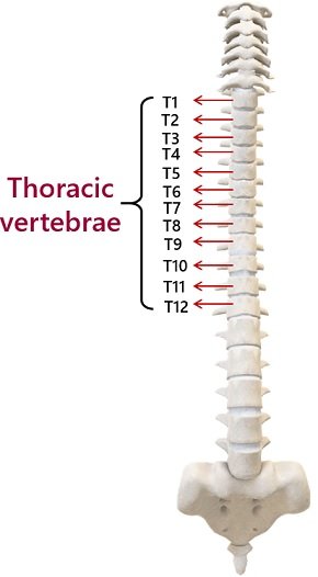
It makes up the second segment of the vertebral spine. The thoracic spine is present between the cervical and lumbar vertebrae. A human body possesses twelve thoracic vertebrae (T1 to T12).
The size of the thoracic vertebra increases downwards. The intervertebral disc is a cartilaginous joint that separates each heart-shaped thoracic vertebra.
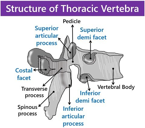
The thoracic vertebra has the vertebral body and the vertebral arch. And, it has the vertebral foramen (hollow space) between the above two components.
Structure
- The superior and inferior view of the vertebral body is heart-shaped. But, the surface seems flat. The vertebral body has a uniform curvature. It comprises two articulation facets (demi facets) near the junction of the pedicle.
- Demi facets are present on each side of the thoracic vertebral body. This region supports the head of the ribs. The superior demi facet (near the root of the pedicle) is larger than the inferior demi facet (near the inferior vertebral notch).
- The pedicle is the rigid bony structure attached superiorly to the vertebral body. The superior notch of the pedicle is narrower than the vertebral notch at the base.
- The vertebral foramen or spinal canal is the large central opening. It supports the structure of the spinal cord.
- Costal facets are present on the tip of transverse processes between the vertebral body and lamina. These depressions articulate with the neck or roughed tubercle of the ribs. Only T1 to T10 vertebrae have costal facets.
- Spinous processes are the elongated portions in the vertebral lamina. This part sharply bends downwards.
- Superior articular processes are flat structures. These project upwards from the junction of the pedicle. Inferior articular processes are intertwined with the lamina. Transverse processes are thick and long that develop from the vertebral arch.
Ribs
They appear as long-curved bones. A human body possesses twelve pairs of ribs that encase the rib cage. In rare cases, some people may have one additional cervical rib.
From the back, ribs attach to the thoracic column. On the contrary, ribs connect to the breast bone via costal cartilage from the front view. The combination of ribs and costal cartilage forms costa.
The costovertebral joint is the junction between the head of ribs and vertebrae. At the costotransverse joint, the rib’s tubercle and the transverse processes of the thoracic vertebra intersect.
Based on the attachment of ribs to the sternum, there are three kinds of ribs:
- True ribs (1-14) attach right through the costal cartilage with the sternum. Ribs and the sternum join at the sternocostal joint. The first rib has a unique feature:
It connects with the clavicle at the costoclavicular joint. - False ribs (15-20) indirectly articulate with the sternum. Their costal cartilages join with the costal cartilage of the seventh rib through the costochondral joint.
- Floating ribs include two distal ribs (21-24). These remain free or do not articulate with the sternum.
Structure
A typical rib structure comprises the head, neck, tubercle and shaft.
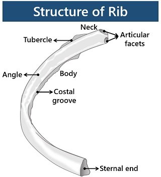
- The head of ribs consists of two articular facets. The superior articular facet connects with the vertebral body’s inferior demi facet. But, the inferior articular facet connects with the vertebral body’s superior demi facet.
- The neck is a flat portion that connects the head to the shaft or body of the rib.
- The roughed tubercle is marked posteriorly between the neck and body. It articulates with the transverse process of the inferior vertebra.
- A rib’s shaft appears thin, and it curves anteriorly by forming a costal angle.
- A costal groove of the body is always pointed downwards.
Thus, ribs are of two types based on the structure.
- Typical ribs (3-9 pairs) have all the components common in their structure.
- Atypical ribs (1,2, 10, 11 and 12 pairs) have some variation. The structure of the first rib comprises two costal grooves and one articular facet. The second rib has a tubercle on the superior region that connects with the serratus anterior muscle. A tenth rib possesses only one articular facet. The eleventh and twelfth ribs lack neck and contain only one articular facet.
Sternum or Breast Bone
It lies in the anterior region of the rib cage and measures a length of 7 cm. The anterior extremity of the sternum is rough and convex. Whereas, the posterior extremity is somewhat concave and smooth.
Structure
The sternum’s typical structure comprises manubrium, body and xiphoid process.
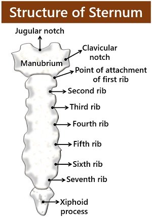
Manubrium or Episternum
- It forms the superior region of the sternum.
- The manubrium appears thicker and broader than the body and xiphoid process.
- Manubrium or episternum has a U-shaped jugular notch.
- Superior lateral margins of the manubrium contain small depressions (clavicular notches).
- It provides a site of attachment between the sternum and clavicle.
- Among twelve pairs of ribs, the first pair attaches to the manubrium.
Body or Mesosternum
- It appears as the elongated central portion.
- The sternal angle forms between the manubrium and the body, as the surface is not flat.
- The second pair of ribs attach to the sternal angle. Yet, a pair of ribs from three to seventh connect to the sternal body.
Xiphoid process or Metasternum
- It is the ossified structure present at the sternum’s inferior tip.
- The xiphoid process is thin and pointed.
Conclusion
Thus, the thoracic cage forms an essential component of the axial skeleton. It anchors organs in the thoracic cavity and supports the upper and lower extremities of the body.