Chlamydia is an obligate intracellular parasite that functions as a “Large virus”. Chlamydias are entirely dependent on the host cells for energy metabolism, like viruses. However, they are true bacteria and differ from viruses by many ways.
Like bacteria, chlamydia also possesses both DNA and RNA. They multiply by binary fission, whereas viruses go through lytic and lysogenic phases in their life cycle. Chlamydia possesses bacterial cell type incorporated with peptidoglycan (probably containing mumaric acid).
They also possess ribosomes, which are absent in viruses. Chlamydiae generally exist as nonmotile coccoids. Chlamydia is the genus comprising three typical species (C. trachomatis, C. psittaci and C. pneumoniae).
In this context, we will study the meaning, features, some unique properties and taxonomy of the genus Chlamydia. Also, the life cycle, pathogenicity, diagnosis, treatment and significance of all the strains of chlamydia have been explained.
Content: Chlamydia
Chlamydia Meaning
Chlamydiae contribute to the small group of gram-negative bacteria, which go through a complex life-cycle or different phases during their intracellular multiplication. They have a variety of metabolically active enzymes. Many antimicrobial drugs may inhibit their growth. Chlamydiae reproduce via binary fission and complete their biphasic developmental cycle within 40-60 hours.
Features of Chlamydia Bacteria
- Gram reaction: They give a negative gram reaction.
- Motility: Chlamydiae are generally non-motile.
- Shape: Chlamydiae are roughly spherical.
- Size: They exist as the smallest prokaryotic organisms whose size ranges between 0.2-1.5 µm.
- Capsule: It is absent in chlamydia.
- Cytoplasmic membrane: It lies under the cell wall and contains high lipid content.
- Genome: The DNA appears as an irregular mass inside the cytoplasm without a nuclear membrane. Chlamydia possesses a genome size of 4-6X10⁸ Daltons and 41-44% of G+C content.
- Ribosomes: These appear as the diffused structures in the cytoplasm.
- Nature: Chlamydiae behave as obligate intracellular parasites.
- Reproduction: Chlamydia reproduces only in the cytoplasmic vesicles of their host’s living cells.
- Chemical nature: They contain high lipid content, especially phospholipid.
- Staining: Chlamydia cells take up the colour of special Giemsa’s stain and Macchiavello’s stain.
Uniqueness
Chlamydiae share some of the unique attributes that make them different from the other gram-negative bacteria.
- Energy parasite: Microbiologists state that Chlamydiae as “Energy parasites” because they lack an ATP generating system but can obtain ATP from the host cell machinery.
- Enzyme system: Chlamydiae comprise few enzymes that aid in synthesizing peptidoglycan. The chlamydial cell wall is devoid of peptidoglycan, but the enzymes synthesizing peptidoglycan accounts for the penicillin effect that disrupts peptidoglycan synthesis and inhibits chlamydial growth.
- FtsZ gene: Chlamydia lack the FtsZ gene, which is necessary for all the bacteria and archaea to form septum during the cell division.
Taxonomy
Based on the pathogenicity and clinical symptoms, there are two ecological groups of chlamydia.
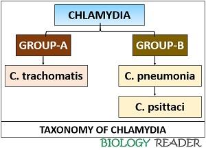
- Group-A includes C. trachomatis that primarily cause trachoma, inclusion conjunctivitis, lymphogranuloma venerum in man only.
- Group-B includes C. pneumonia and C. psittaci that mainly infect birds, which can be transmitted to men and result in zoonotic infections.
Life Cycle of Chlamydia
Chlamydia possesses a very complex growth and multiplication cycle, where it goes through different phases. The growth, reproduction and maturation of chlamydia usually take 48 hours. Its life cycle exists between two morphologically distinct forms or cell types.
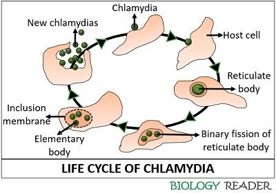
- Elementary bodies or Chlamydiospores exist as small, rigid-walled, infectious particles that remain alive even after their exit from the host cells.
- Reticulate or Initial bodies appear larger, thin-walled, non-infectious particles that undergo cell division via binary fission.
| Properties | Elementary body | Reticulate body |
|---|---|---|
| Size | 0.3 µ | 0.5-1.0 µ |
| RNA to DNA ratio | 1:1 | 3:1 |
| Pathogenicity | Infectious | Non-infectious |
| Role | Adapted for extracellular survival | Adapted for intracellular growth |
| Haemagglutinin | Present | Absent |
| Endocytosis | It can induce endocytosis | It does not |
| Metabolic activity | It is metabolically inactive | It is metabolically active |
Stages of Growth and Multiplication
Chlamydia goes through a series of developmental stages, as summarized below:
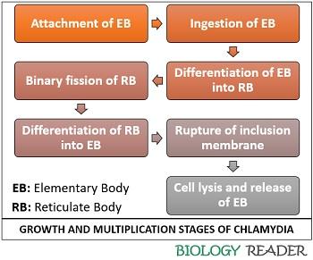
- Attachment: Firstly, the small cell types or elementary bodies (highly infectious) attach to the cell surface of a host cell and enters inside it.
- Ingestion: Then, a host cell ingests the elementary bodies through an endocytic process (Phagocytosis).
- Differentiation: Once the elementary bodies enter a host cell, they retain their structural integrity inside the membrane-bound vesicles. During this stage, there is no eclipse (where a cell loses its infectious ability). In this stage, the small cell types or elementary bodies morphologically change into large cell types or reticulate bodies.
- Cell division: The reticulate bodies continuously enlarge in size and undergo repeated multiplication by binary fission after (10-15 hours) of infection.
- Differentiation: Afterwards, the reticulate bodies tend to decrease and transform into typical elementary bodies to complete the developmental cycle. This stage involves the reorganization of the large cell types into small ones.
- Cell lysis: The infected host cell undergoes cell lysis after 42 hours of infection, which results in rupturing of the inclusion membrane.
- Host cell lysis: Finally, the host cell ruptures after 48 hours of infection and eventually releases infectious elementary bodies that later infect new healthy cells.
Pathogenicity
- C. trachomatis predominantly affects the mucus membrane of the eye and genitourinary tract of humans.
- C. pneumoniae and C. psittaci primarily infect the bird (carriers of the infection), which further cause the dissemination of the infection in men. An infected person may acquire various respiratory illnesses.
Diagnosis
A person diagnosed with infections by the Chlamydia strains needs to be tested by the following serological tests:
- Microimmunoflourescent test can be employed for patients with an eye infection.
- The direct immunofluorescent test can be employed for patients suffering from neonatal conjunctivitis and early trachoma.
- Frei test (type IV hypersensitivity test) used to kill organisms in the genitourinary tract.
Treatment
The patients infected with C. trachomatis can be treated by antibiotics (doxycycline or azithromycin). Doxycycline or azithromycin, or erythromycin, can be given to the patients infected with C. pneumoniae, while doxycycline and erythromycin can be given to those infected with C. psittaci.
The diseases caused by chlamydiae are relatively easy to treat but have two problems:
- Latency of infection: The infection may remain latent or sub-clinical for years.
- The susceptibility of the compromised host to reinfection.
Significance
C trachomatis, C. psittaci and C. pneumoniae are the only species of the genus chlamydia recognized so far. C trachomatis is the causative agent of two prevalent human diseases; the genitourinary tract disease (Lymphogranuloma venerum) and the conjunctivitis infection (trachoma).
C. psittaci causes psittacosis disease in birds, which occasionally transmitted to humans and caused pneumonia-like symptoms. C. pneumoniae is the causal organism associated with a variety of respiratory syndromes in humans.