The difference between apoptosis and necrosis is mainly due to factors like:
Cell death: Apoptosis is a type of cell death, which occurs by the self-annihilation of the cell. Oppositely, necrosis is a form of focal cell death caused due to the environmental perturbations.
Occurrence: Apoptosis occurs to maintain the proper functioning of the cell by removing the dead or metabolically inactive cells via phagocytosis. In contrast, necrosis occurs as a result of cell exposure to cytotoxic agents.
Content: Apoptosis Vs Necrosis
- Comparison Chart
- Definition
- Characteristic Events
- Cellular Changes
- Types
- Key Differences
- Similarities
- Conclusion
Comparison Chart
| Properties | Apoptosis | Necrosis |
|---|---|---|
| Meaning | It is a natural process of cell death, which occurs inside the body and regulated by the cell itself | It is an accidental cell death, which occurs due to exposure of the cell to some harmful physical, chemical and biological agents |
| Alternative names | It can be interchangeable with terms like cell suicide, shrinkage necrosis or active or autophagic cell death | It can be interchangeable with terms like accidental cell death or passive cell death |
| Form of cell death | It is predefined or programmed cell death | It is unexpected or accidental cell death |
| Cause | It occurs naturally by the self-annihilation of the cells | It occurs in response to trauma, ischemia, excitotoxicity, infection, hypoxia etc. |
| Regulation | It is a regulated process that is genetically controlled | It is an unregulated process that is not regulated genetically |
| Energy (ATP) input | It is ATP-dependent | It is ATP-independent |
| Process | It is a physiological process mediated by the cells itself | It is a pathological process induced by external factors like toxins, drugs etc. |
| Caspase dependency | Apoptosis is a caspase-dependent pathway | It is caspase-independent pathway |
| Vesicle formation | Apoptotic bodies are formed in apoptosis, which is nothing but the small fragments of the cell | Necrosis does not involve any vesicle formation, as the complete cell undergoes cell lysis |
| Release of protein factors | The mitochondria of apoptotic cell release mitochondrial factors like Cyt-c and AIF into the cytosol | This process does not release any protein factors |
| Affects | It affects the individual cells or small groups of the cell | It affects large contiguous cell groups |
| Cellular change | Cell shrinkage | Cell swelling |
| Phagocytosis of cell remnants | Apoptotic bodies are phagocytized by the specialized macrophages or by the adjacent cells | Necrotic cells are phagocytized by macrophages only |
| DNA electrophoresis | Step ladder pattern | Smear pattern |
| Death inducing stimulus | Less severe and transient stresses induce apoptosis | Severe and sustained stresses induce necrosis |
| Eliciting inflammatory response | It does not elicit an inflammatory response | It triggers an inflammatory response |
| Symptoms | No visible symptoms | Necrosis generally causes acute inflammation, tissue damage, scar formation etc. |
| Treatment | It does not require any clinical treatment | It requires clinical treatment, as untreated necrosis can be fatal or detrimental |
| Enzymes involved in cell degradation | Caspases are the key enzymes that mediate cell proteolysis | Calpains, cathepsins B and D are the enzymes involved in the cell proteolysis |
| Effect on the cell organelles | ||
| Plasma membrane | It remains intact | It gets disrupted |
| Mitochondria | It becomes leaky | Not affected |
| Lysosomes | Not affected | It becomes leaky |
| Chromatin | Chromatin becomes condensed or aggregated | Chromatin becomes fragmented |
| Nucleus | It gets fragmented | It gets disorganized |
| DNA fragmentation | Pre-lytic DNA fragmentation occurs in the internucleosomal region | Post-lytic DNA fragmentation occurs |
Definition of Apoptosis
Apoptosis can define as the innate process of cell death, which is generally regulated by the cells itself to maintain the cell number (ratio of viable and dead cells). It does not cause any harm to the tissues, but its abnormal functioning may impose some severe health effects.
Apoptosis is a caspase-dependent cell death pathway that mediates membrane blebbing, cell fragmentation and cell proteolysis. Caspases include a large group of proteases, which participates in the predefine cell death process. Upon activation of caspase, the scaffold proteins (lamins) cleave that results in nuclear shrinkage and fragmentation.
Plasma membrane blebbing occurs due to the breakdown of gelsolin by the caspase-3 enzyme. A cytoskeleton protein (Fodrin) is also cleaved by caspases, which results in an overall loss of cell integrity, size or shape.
Definition of Necrosis
Necrosis can define the kind of accidental cell death, which is not regulated by the cells. It is induced by an overdose of cytotoxic factors like drugs, toxin, ischemia etc. or environment perturbations like radiation, lack of oxygen (hypoxia) etc.
Necrosis is a passive and degenerative process, which do not need ATP. It triggers the inflammatory response in the abnormal cells or diseased organ or tissue as a result of cytolysis. It often results in scar formation along with inflammation.
Necrosis is a form of focal or localized cell death, which occurs in the particular cells or tissues affected by some cytotoxic agents. During necrosis, swelling of the cell is the first event, which is later followed by the blebbing of the plasma membrane and cytolysis.
Characteristic Events
Apoptosis: It involves a series of events like:
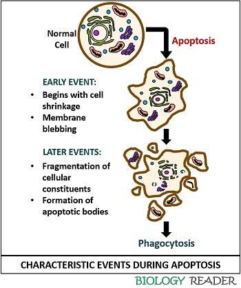
Early events:
- A cell becomes shrivelled.
- Membrane blebbing occurs
Later events:
- Cytoplasmic calcium concentration increases
- Increased lipid peroxidation
- Pyknosis occurs (Chromatin condensation towards nuclear periphery)
- Proteolysis by caspase
- Fragmentation of nucleus (Karorrhexis) and cell
- Formation of apoptotic bodies
- Phagocytosis by the professional macrophages or adjacent cells
Necrosis: It includes a series of events like:
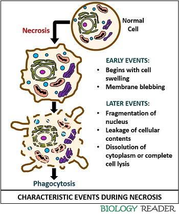
Early events:
- Dysregulation of ion homeostasis
- Cell becomes turgid
- Membrane blebbing occurs
Later events:
- Chromatin fragmentation or Pyknosis occurs.
- Nucleus fragmentation (Karorrhexis)
- Swelling of cell mitochondria
- Rupture of plasma membrane
- Dissolution of cytoplasm or cytolysis takes place.
- Release of cytoplasmic constituents
- Phagocytosis of cytoplasmic contents by the professional macrophages
Cellular Changes
Let us discuss the morphological, biochemical, physiological and molecular changes in the cells during apoptosis and necrosis.
Morphological Changes
Apoptosis causes membrane blebbing and margination, without affecting the membrane integrity. The chromatin material of apoptotic cell becomes condensed due to the cellular shrinkage. It also forms persistent apoptotic bodies.
Necrosis cause membrane blebbing and margination, which loosens the membrane integrity. The chromatin material of necrotic cell becomes randomly fragmented due to the increase in cell size. It causes cell-lysis and disintegration of cellular contents.
Biochemical Changes
Apoptosis process is genetically controlled and resulting in the formation of non-random oligonucleosomes of DNA. It is an active cell death process, which requires energy input or ATP. In apoptosis, pre-lytic DNA fragmentation occurs.
The loss of ion homeostasis marks biochemical changes. It is a passive cell death process, which does not require energy input or ATP. In necrosis, post-lytic DNA fragmentation occurs.
Physiological Changes
Apoptosis causes the death of either a single cell or small group of cell, which is evoked by the physiological stimuli (by the cell itself). The apoptotic bodies are removed by the specialized macrophages or by the neighbouring cells. Apoptosis does not cause tissue damage and inflammation.
Necrosis causes the death of a large group of adjacent cells, which is evoked by the pathological stimuli (by the external factors like temperature, chemicals etc.). The specialized macrophages eliminate the necrotic cells. Necrosis causes tissue damage and acute inflammation.
Molecular Changes
In apoptosis, cell degradation and fragmentation is triggered by the activation of the caspase enzyme. Cell apoptosis occurs due to the loss of normal cell survival signals or by the action of harmful agents.
In necrosis, cell degradation and fragmentation occurs by the release of hydrolytic enzymes after the breakdown of lysosomes. Cell necrosis occurs due to harmful chemical, physical or chemical agents. It occurs due to the ATP depletion, membrane damage and free radical injury.
Types
Apoptosis: It generally includes two types of cell death pathway, namely intrinsic and extrinsic cell death pathway.
- Intrinsic apoptosis: In this type, a cell receives a signal from its genes to kill itself once it senses any cell stress. Intrinsic cell death is a mediated by the mitochondrial proteins, which mainly involves BAX and BAK heterodimers. It results in the development of pores in mitochondria, which then releases cytochrome-c and DIABLO proteins to activate extracellular caspases.
- Extrinsic apoptosis: Here, the cell receives a signal from the other cells to undergo cell death. It is a receptor-mediated process, which involves death receptors (TNF and Fas) that activates the intracellular caspases and dependence receptors (DCC and PTCH1). Both types involve caspase activation for cellular degradation.
Necrosis: It is mainly of six types:
- Coagulative: It is caused due to severe ischemia.
- Liquefactive or colliquative: It is caused due to bacterial or fungal infection.
- Gangrenous: It is caused due to ischemia of the lower limbs.
- Caseous: It is caused due to mycobacteria, fungi or some other antigens.
- Fibrinoid: It is caused by immune-mediated vascular damage.
- Fat necrosis: It is caused by the necrosis in the fatty tissues.
Key Differences Between Apoptosis and Necrosis
- Apoptosis is a natural cell death induced physiologically by the cells itself, and it is a predefined or regulated cell death, which is controlled genetically. Oppositely, necrosis is an accidental cell death induced by an overdose of cytotoxic agents, and it is a kind of undefined cell death that can cause localized damage to the cells or tissues.
- Apoptosis is an active process, which generally occurs in the individual cells and requires energy input in the form of ATP. In contrast, necrosis is a passive and degenerative process that may occur in the contiguous group of cells and do not need energy input.
- Caspases are a large group of protease enzymes, which facilitate cell degradation or cell proteolysis in the type of programmed cell death, i.e. apoptosis. Therefore, apoptosis is caspase-dependent and necrosis is caspase-independent.
- The early event in apoptosis and necrosis also provides a characteristic difference between the two, where the initial one undergoes cell shrinkage and the latter undergoes cell swelling.
- Membrane integrity is one of the striking difference between the apoptosis and necrosis, in which a former do not lose membrane integrity, i.e. the membrane remains intact, and the latter loses the membrane integrity, i.e. the plasma membrane ruptures.
- In apoptosis, the cell undergoes vesicle formation or the formation of apoptotic bodies that are later phagocytosed by the specialized macrophages or the neighbouring cells. In necrosis, a cell does not undergo vesicle formation, and the cell contents leak out in the extracellular matrix that is later phagocytized by the professional macrophages.
Similarities
- Apoptosis and necrosis are the two different mechanisms of cell death.
- Both the processes are common in eukaryotes.
- Nucleus fragmentation or Karorrhexis occurs in both apoptosis and necrosis.
- Both the apoptotic bodies and the cell remnants of necrotic cells go through phagocytosis.
Conclusion
Therefore, we can conclude that the apoptosis and necrosis are both different kinds of cell death that involves different mechanism, events etc. But, both apoptosis and necrosis are the cell death processes marked by profound membrane dysfunction.