Haemagglutination is a kind of agglutination reaction that includes erythrocytes. The result is characterized by the agglutination or clumping of erythrocytes. Haemagglutination can be assayed in blood typing and viral quantification.
The reason behind the haemagglutination reaction is the glycoproteins that cause RBCs to agglutinate and serves as “Hemagglutinins”. Antibodies and lectins are commonly known as haemagglutinins.
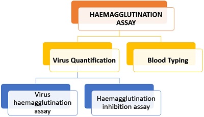
Content: Haemagglutination
Meaning
Haemagglutination can be defined by breaking the term into two terms, i.e. haem that is the part of RBCs and agglutination means an aggregation of particles. Thus, haemagglutination reaction refers to the reaction that facilitates clumping of RBCs via the agglutinating agents or agglutinins. It is commonly used in blood typing to examine the known surface antigens on RBCs. In addition to this, it also quantifies the presence of unknown antigens like bacteria, viruses etc. The result of the haemagglutination test can be seen macroscopically.
Haemagglutination Assay
Haemagglutination can be used to screen for antibodies in RBCs (with known surface antigens) in blood typing. It can also be employed for viral quantification by preparing the serial dilutions of virus suspension.
Blood Typing
In ABO blood typing, the haemagglutination reaction confirms the type of antibodies (either anti-A and anti-B antibodies or both), which mainly binds to the surface antigens (either A, B or both). The binding of surface antigens on erythrocytes with the corresponding antibodies results in haemagglutination or clumping of RBCs.
To perform blood grouping, we need to take a small sample of blood into the well of the haemocytometer slide. Then add antiserum (A, B and D) to the blood sample, and allow the slide to stand for a few minutes.
If agglutination occurs in the blood sample added with antiserum-A, then the person has a B-blood type. Similarly, if agglutination of RBCs occurs in the blood sample with antisera-B, then the person has an A-blood type. If haemagglutination occurs in the blood sample added with antisera- A and antisera-B, then the person has AB-blood type. If there is no haemagglutination, then the person has an O-blood type. The addition of antisera-D is added to know the Rhesus status of the individual, whether a person is Rh-positive (agglutination occurs) or negative.
Viral Quantification
Viral haemagglutination assay involves serial dilution of virus suspension followed by the sequential steps that are as follows:
- First, prepare virus suspension and then perform serial dilution in the successive wells in a microtiter plate. An assay tray or microtiter plate comprises a series of shallow wells of uniform volume.
- Then add a standard amount of RBCs.
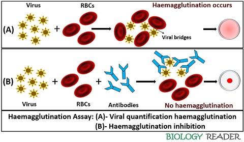
- The viral antigens possess some protein through which these can agglutinate the erythrocytes, thus preventing them from settling out of suspension.
The less diluted HA titre will contain more cells that will attach to more than one RBC, and cause agglutination. In contrast, the more diluted suspension will contain fewer numbers or fewer viruses and cannot promote the clumping of blood cells. This method is cheaper and quicker.
The positive result indicates the formation of diffused mat over the round-bottomed wells. In contrast, the negative result will show the absence of viral particles and appear in the form of sediment in the centre of the shallow wells. The concentration of antigen or virus can be measured by a haemagglutination factor that contains the minimum number of viruses that results in complete agglutination of erythrocytes.
Haemagglutination Inhibition Assay
The addition of an antiserum can modify the viral haemagglutination assay. It involves the addition of a standard amount of virus into the wells of the microtiter tray. Then serially diluted antiserum is added over the viral suspension. At last, RBCs is added in an equal proportion to each well. The microtiter tray is then left undisturbed for at least 30 minutes, after which the readings can be noted.
More concentration of antiserum contains more antibodies that will compete more with the viral cells from getting attached to the RBCs. In contrast, the more diluted antiserum will contain less or no antibodies, which can result in haemagglutination. The result in HIA can be interpreted when the lowest concentration of antiserum inhibits haemagglutination or forms clear sediment at the bottom of the microtiter plate.
Advantages
- Haemagglutination assay is a simple and reliable method to know the concentration of surface antigens on the RBCs and the particulate antigens.
- It is a relatively inexpensive assay that gives results within a few hours.
- The assays allow a measure of comparison and standardization.
Limitations
- To obtain reliable results, technical skills are required who having much idea over the parameters like incubation time, concentration and type of erythrocytes etc.
- Non-specific factors may interfere with the titre values and can give incorrect results.
Conclusion
Therefore, we can conclude that the haemagglutination reaction merely involves the participation of RBCs, where these get agglutinated by the protein factor-like haemagglutinins.