Human skin layers constitute the body’s outer covering that shields the internal cells, tissues, and organs against the changing environment, allergens, and pathogens. The human skin is the largest organ among the other components of the integumentary system. Besides its immunity role, skin regulates body temperature, synthesis of vitamin D, and sensation of touch, heat, pressure etc.
The skin protects the body against UV damage, excessive water loss and foreign bodies like bacteria. Generally, two layers of skin (namely, epidermis and dermis) are connected to the blood vessels and underlying bones and muscles through a subcutis or hypodermis layer.
Thus, the epidermis is the skin’s outermost layer comprising five sublayers starting from the basement membrane (stratum basale, stratum spinosum, stratum granulosum, stratum lucidum and stratum corneum) along with specialized cells like Merkel cells, melanocytes, Langerhans cells and keratinocytes.
The dermis is beneath the epidermis layer, comprising a pilosebaceous unit, fibroblasts, collagen, mast cells and capillaries. Subcutis is the innermost layer containing loose connective tissues, fatty tissues and elastin, which acts as an intermediate link between the skin and bones.
Content: Human Skin Layers
Definition of Skin
The skin is a part of an integumentary system forming the outer covering of the organisms, which protects the cell’s interior against dehydration, abrasion, invasion of microbes, and physical and chemical stresses. Broadly, skin can be classified into glabrous (hairless) and hairy types. Besides the skin, appendages like hairs and nails are also components of the human integumentary system.

Human skin appears as a thick layer of keratinized epithelium that comprises five sublayers and a dermis layer below it. Moreover, you will get to know all the components of the epidermis and dermis layers of the human skin, along with their functions and some important facts.
Anatomy of Human Skin
Human skin majorly comprises two layers (epidermis and dermis), but a layer that connects the skin to the bone is known as hypodermis (not a part of the skin). Here, we will briefly discuss each functional component of the epidermis and dermis layer of the human skin, along with the diagrammatic representations.
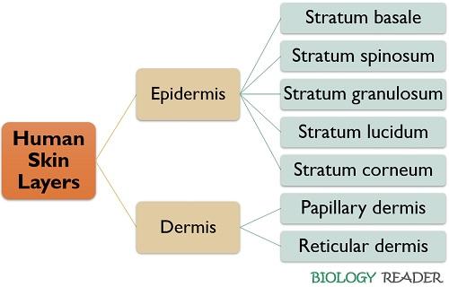
Epidermis
It is the topmost layer of the human skin that possesses five sublayers.
- Stratum basale: It is the first layer above the basement membrane possessing column-shaped basal cells. The old basal cells shift upwards to the squamous cell layer. The basal cells get flattened and die once they become old. It possesses one receptor cell called the Merkel cell that responds to the pressure exerted on the skin.
- Stratum spinosum: It possesses flattened cells, Langerhans cells and specialized melanocytes. The Langerhans cells are the dendritic cells that provide primary defence against microbial invasion. Melanocytes synthesize melanin pigment and control the skin tone (the higher the melanin production, the darker your skin colour). Melanosome vesicles act as a packaging material that stores melanin, and their number and size determine the skin tone.
- Stratum granulosum: It contains more keratinocytes that synthesize a colourless protein (keratin). The old keratinocytes will move upwards (towards the skin’s surface). Keratin strengthens the skin.
- Stratum lucidum: It only exists in the thick skin (soles, palms etc.).
- Stratum corneum: This layer is the thickest and outermost squamous cell layer, which comprises dead and flattened corneocytes (lack nucleus). It sheds off the dead keratinocytes (desquamation) after every 28-30 days and replaces them with new keratinocytes.
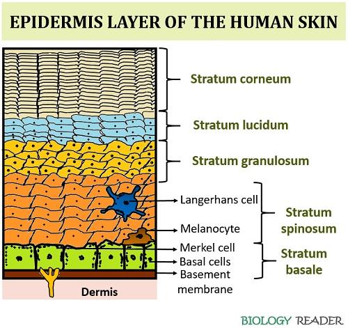
The epidermis layer possesses four specialized cells:
- Merkel cells: It functions as receptor cell that responds to pressure.
- Melanocytes: These make the melanin pigment that decides the human skin’s tone.
- Langerhans cells: These are the dendritic cells forming the first line of defence against the outside environment.
- Keratinocytes: These form colourless protein keratin that provides strength.
Dermis
It is an intermediate layer between the basement membrane and the subcutis. It has two sub-layers, namely the papillary and reticular dermis. The dermis layer is the thickest portion of the skin that constitutes about 90% of the human skin. It comprises most of the specialized cells and structures. A papillary dermis is present underneath the basement membrane, while a reticular region is associated with the lower region of the hypodermis layer.
- The papillary layer appears thin and comprises loose connective tissues. It possesses some irregular projections called papillae or epidermis ridges. It includes an extensive network of blood vessels that supplies essential nutrients to the epidermis layer and supports keratinocytes’ formation.
- The reticular layer appears thick and contains compactly packed or irregular connective tissues. It possesses collagenous, elastic and reticular fibres along with the pilosebaceous unit. Collagen (mostly type-1 and 3) and elastin proteins are the components constituting the formation of the reticular layer, in which the former provides strength and the latter gives elasticity to the human skin.
Let us discuss the specialized cells and structures that exist in the skin’s dermis layer:
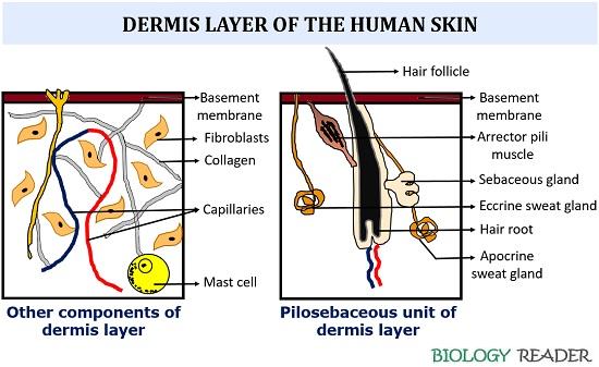
Pilosebaceous Unit
It comprises hair follicle, arrector pili muscle, sebaceous gland and sweat gland, colloquially termed epidermal invagination.
- Hair follicle: It originates from the follicular base called the hair bulb. Each hair follicle contains different cells and connective tissues. The living cells in the dermis layer’s hair bulb will divide actively to build a hair shaft. The network of blood vessels nourishes the cells dividing in the hair bulb.
- Arrector pili muscle: It is composed of smooth muscle fibres and attached to a follicular bulge. Contraction of this muscle results in piloerection or goosebumps.
- Sebaceous gland: It is an exocrine gland that secretes oil into the skin. The sebaceous gland is associated with the hair follicle. It functions as a lubricating gland of the skin that maintains the body’s moisture. Oil glands are absent in the palms and soles.
- Sweat gland: It is also called the sudoriferous gland. Apocrine and eccrine are the two common sweat glands that differ in location, function and secretory product. The apocrine sweat gland is found in the hairy skin (armpits, groin area etc.), and the eccrine sweat gland is located all over the body. The apocrine and eccrine gland’s primary function is to contribute to body odour and regulate the body’s temperature, respectively. Numerous adipose tissues contribute to the lining of sweat glands.
Mechanoreceptors & Specialized Cells
Pacinian and Meissner corpuscles are the cutaneous receptors which respond to the vibratory sensation. Skin includes specialized cells like fibroblasts, macrophages, adipocytes, and mast cells. Fibroblasts are the specialized cells responsible for the production of collagen, elastic and reticular fibres along with the extracellular matrix. Macrophages (histiocytes) assist the immune system. Adipocytes provide insulation and hair follicle regeneration. Mast cells generate an inflammatory response.
Subcutis
It is not a part of the skin but is found to be associated with the lower region of the dermis layer. It is also called the subcutaneous and hypodermis layer. Subcutis allows the components of the skin to communicate with the underlying fibrous tissues of bones and muscles. A network of loose connective tissues, fat cells and elastin proteins contribute to the subcutis layer.
Video: Anatomy and Physiology of Human Skin
Functions of Human Skin
Skin performs various functions:
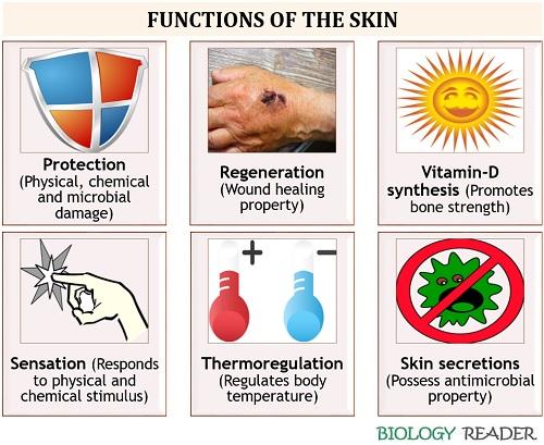
Protection
Skin shields the body and provides primary defence against physical, chemical and biological damage. Also, it prevents excessive water loss or maintains body homeostasis.
Regeneration
Skin can regenerate new cells by replacing the old, dead or damaged cells. Skin regeneration is one of the skin’s important features, as the skin cells renew after 28-30 days. The dead cells from the lower epidermis reach the skin’s surface, where they grow hard and sloughed off from the skin.
Vitamin-D Synthesis
Skin functions as a site of vitamin-D synthesis, in which the substance (7-dehydrocholestrol) isomerizes into cholecalciferol or previtamin-D3 in the presence of UVB light. This inactive form (cholecalciferol) further hydroxylates in the liver and kidney to produce an active form of vitamin D (calcitriol). In the bloodstream, calcitriol travels as a hormone and functions to regulate calcium and phosphate synthesis, promoting bones’ remodelling.
Sensation
Human skin possesses mechanoreceptors, thermoreceptors and nociceptors that can sense the pain exerted by deep pressure, temperature and noxious stimuli.
Thermoregulation
Thermoregulation is one of the skin’s major functions, which can either occur through blood vessels or sweat glands. The blood vessels dissipate heat from the skin as the body temperature increases through vasodilation. Oppositely, the blood vessels retain heat as the body temperature decreases through vasoconstriction. Similarly, the sweat glands in the skin’s dermis layer transpirate heat out by releasing electrolytes out of the skin through sweating or perspiration.
Secretion
The skin’s secretions, like sebum (from sebaceous glands) and sweat (from sweat glands), act as antimicrobial agents. Sebum is the waxy lipid material that lubricates the skin that forms a slightly acidic environment to restrict the direct contact of microorganisms like bacteria, fungi etc. Similarly, the sweat glands release electrolytes (NaCl) that also inhibit microbial interaction.
| Properties | Epidermis | Dermis | Subcutis |
|---|---|---|---|
| Composition | It is composed of stratified epithelium, keratinocytes, melanocytes and Langerhans cells | It is composed of the pilosebaceous unit, specialized cells and mechanoreceptors | It is made of subcutaneous fat |
| Functions | Immune defence, decides skin tone, protects against uv-damage and pathogens | Pliability, thermoregulation, tensile strength, wound-healing and elasticity | Insulation, calorie reserve and shock absorber |
Fun Facts about the Human Skin
- It’s the largest organ of the human integumentary system, but the small intestine is the largest organ found in our body’s interior.
- The skin comprises 19 million skin cells.
- There are about 20 blood vessels, and 1000 nerve endings exist within the skin.
- The skin possesses approximately 650 sweat glands.
- Skin contributes approximately 12-15% of the body’s weight.
- The skin forms the first line of defence against physical, chemical and biological damage.
- Skin thickness varies accordingly to the different body parts and depends on the person’s age, gender and eating habits. Our feet have a maximum thickness (1.4mm), while our eyelids possess the thinnest skin (0.2mm).
- The skin tone is decided by the melanin pigment released by the melanocytes in the epidermis layer. The more melanin pigment, the darker the colour of your skin.
- Legs contain a few oil glands, due to which they are considered the driest body part.
Conclusion
Therefore, we can conclude that the human skin possesses an epidermis and dermis layer where each performs distinct roles. Skin performs multitasking, as it acts as a barrier or shock absorber by sensing the physical or chemical (UV-light, physical damage and trauma etc.), maintains equilibrium between the body fluids, regulates the cooling effect in summers, and replaces the old or dead cells. So, we must keep our skin healthy by avoiding excessive UV exposure, unhealthy diet, and stress.