The structure of paramecium seems slipper-like. Paramecium comprises organelles common to eukaryotes. They contain specialized membrane-bound organelles and a well-defined nucleus. Besides that, they are unique in having contractile vacuoles, mouth pore and anal pore.
Paramecium contains organelles necessary to sustain life, including growth, metabolism and reproduction. For the movement, they have small hair-like outgrowths or cilia.
The pellicle is the stiff outer covering that maintains the paramecium’s shape. For osmoregulation, paramecium species have contractile vacuoles.
They have an oral groove to ingest the surrounding food material. For food digestion, the paramecium comprises vesicles containing hydrolytic enzymes. Paramecium species also have cytoproct as a defecation site to excrete wastes.
For the defence against predators, they have trichocysts. Cell organelle like mitochondria helps in respiration and energy production. This post describes the paramecium’s size, appearance, and structure in detail.
Content: Structure of Paramecium
Size
Members of the genus Paramecium are microscopic but visible to the naked eye. Their size differs from species to species.
- Length ranges between 80 to 350 µ.
- The diameter ranges from 170 to 290 µ.
Appearance
Paramecium species fall into two groups based on appearance.
1. Aurelia Group
- Paramecium species belonging to the Aurelia group have an oblong or cigar-shaped body.
- In the cross-section, the body appears round with a tapered posterior end.
- Species like P. aurelia, P. caudatum, P. multimicronucleatum etc., belong to this group.
- This group has sub-terminal cytoproct.
2. Bursaria Group
- Paramecium species belonging to this group appear slipper-like or foot imprints.
- They have a shorter length with a broader body.
- Species of the Bursaria group have a flat body on both surfaces.
- Their body has a rounded posterior end and truncated anterior end.
- Species like P. bursaria, P. calkinsi, P. woodruffi etc., are part of this group.
- This group has a terminal cytoproct.
Paramecium Structure
The structure of paramecium consists of the following components:
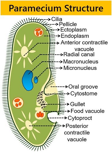
Pellicle
It is a stiff, although elastic membrane surrounding the paramecium body. Pellicle or Periplast consists of a gelatinous substance. It maintains the definite shape of the paramecium.
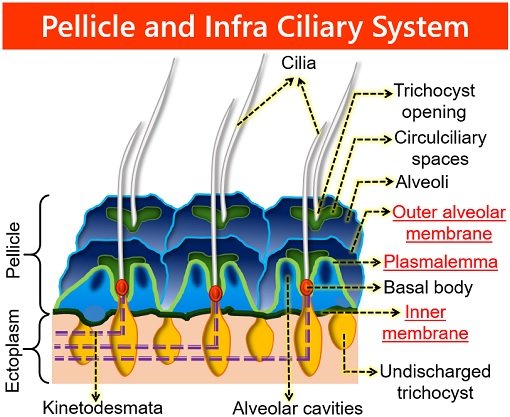
Although, a pellicle is flexible enough to allow paramecium to change its shape. Also, it protects the inner cellular contents. A pellicle consists of three membranes:
- Outer plasmalemma or cell membrane
- Flattened membrane-bound sacs or outer alveolar membrane
- Inner membrane or epiplasm
The outer membranes are continuous with the cilia. And the inner membrane holds the ectoplasm. The pellicle is not smooth (comprises regular series of hexagonal or rectangular depressions).
Each hexagonal depression has a perforation called a central aperture. From a central aperture, a cilium projects out. The hexagonal depressions bear the openings of trichocysts.
Some Paramecium species have spindle-shaped trichocysts. These explosive organelles have a defensive mechanism by discharging thin, non-toxic filaments.
Cilia
Many tiny hair-like cilia are present throughout the pellicle. Their number and arrangement vary from species to species. In approx, a cilium has 0.25 μm diameter and 20 μm length. A body of paramecium contains oral cilia and body cilia.
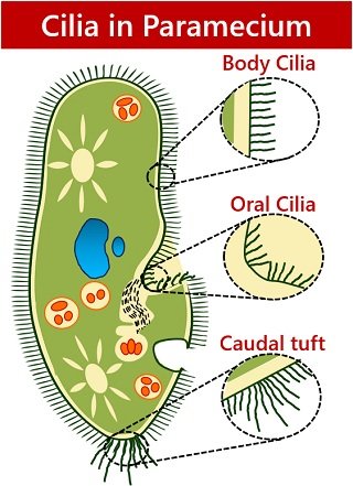
- Body cilia encircle the body surface. They have a uniform length and longitudinal arrangement. Some species have several longer cilia on the posterior end as a caudal tuft of cilia. Body cilia help in the locomotion of the organism.
- Oral cilia surround the surface of the oral groove. They bring the food inbound to the water current into the oral groove.
- A caudal tuft of cilia is tactile or sensitive. Some P. species have several longer cilia at the posterior end in the form of a bunch.
Ultrastructure of Cilia: Like flagella, cilia also have fibrillar composition. Unlike flagella, cilia are present various in number with shorter sizes. The ciliary membrane has a thickness of 90 Å. The liquid matrix of cilia comprises:
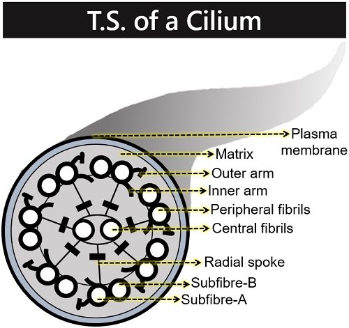
- Nine double longitudinal peripheral fibrils. Here, each ring has two sub-fibrils (A and B).
- Two longitudinal central fibrils. They lack paired sub-fibrils, but each contains a single tubule. A sheath holds these two longitudinal fibrils and gives out nine radial spokes.
A cilium originates from the kinetosome. Besides locomotion and food capturing, cilia can sense external stimuli. Also, the infra-ciliary system helps in forming cell organelles during cell division.
Oral Apparatus
It comprises the following structures:
Oral Groove
It is a large oblique shallow depression on the ventral surface of the paramecium’s body. The oral groove is responsible for the asymmetrical shape of the paramecium.
The oral groove allows the entry of food materials into the cell using oral cilia. It is present in the middle. From the lower end, an oral groove forms a conical vestibule. This complex is in touch with the wide tubular passage, i.e. the buccal cavity.
It then leads to the wide cytopharynx through a mouth pore or cytostome. The cytopharynx produces a spherical food vacuole at the proximal end.
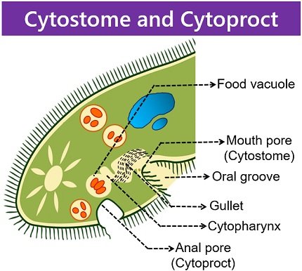
Cytostome
It is the mouth pore that appears funnel-like. Oral cilia also cover the lumen of the cytostome. Here, the oral cilia bring the food material down into the cytopharynx. The tubular cytopharynx forms food vacuoles through budding.
Cytoproct or Anal Pore
Paramecia excrete the undigested waste after nutrient absorption. It is present on the ventral surface. Food vacuoles containing indigestible food fuse with the pellicle to release the debris. The anal pore of paramecia does not have ridges and cilia.
Cytoplasm
It comprises two distinct areas, i.e. ectoplasm and endoplasm. Ectoplasm is a thin, firm outer layer anchoring the roots of cilia. Also, it comprises trichocysts and fibrillar components. Ectoplasm plays a major role in protecting the paramecium body against damage.
Endoplasm is the metabolically active region holding different cell organelles. It comprises all organelles of the eukaryotic cells like:
- Mitochondria (the powerhouse of the cell)
- Endoplasmic reticulum and ribosome (Involved in protein synthesis)
- Golgi apparatus (helps in transporting, processing and packaging of proteins)
- Lysosomes (holds digestion enzymes)
Besides, paramecium contains two nuclei, food and contractile vacuoles.
Nucleus
Paramecium has a macro and micronucleus.
1. Macronucleus: It appears ellipsoidal or kidney-like with dense chromatin granules (DNA). It is also called a generative or vegetative nucleus and present one in number.
The macronucleus regulates vegetative or non-reproductive cell functions. It has a subset of DNA from the micronucleus. It is polyploid or contains up to 800 copies of the chromosome.
2. Micronucleus: It exists as small, compact and spherical. It is present close to the macronucleus, and the number can vary from species to species. The micronucleus is diploid and contains all the DNA materials.
It controls cell reproduction. Based on the type of micronucleus, the members of paramecium fall into two specific types:
- Caudatum type: P. caudatum, P. bursaria, P. trichium etc., have large and ellipsoidal micronucleus. They comprise a compact mass of chromatin within the micronucleus. We could see the nuclear membrane around the micronucleus using a light microscope.
- Aurelia type: P. aurelia, P. woodruffi, P. jenningsi etc., have small and vesicular micronucleus. They comprise endosomes with a less concentrated mass of chromatin. Here also, you could see the nuclear membrane surrounding the micronucleus.
Food Vacuoles
These are non-contractile and roughly spherical. First, a paramecium captures the food from the oral groove to the cytopharynx via a mouth pore.
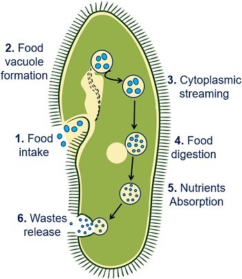
Then, the budding of the gullet gives rise to the formation of food vacuoles. These function like the temporary stomach. Food vacuoles fuse with the lysosomes in the cytoplasm.
The digestive enzymes within lysosomes break down food materials into simpler nutrients. The size of food vacuoles varies depending on food intake and digestion. A paramecium cell ejects the cell debris outside the body through the anal pore.
Contractile Vacuoles
These appear star-shaped. Single paramecium cell has contractile vacuoles at the anterior or posterior ends. They regulate osmoregulation and excretion of water and wastes.
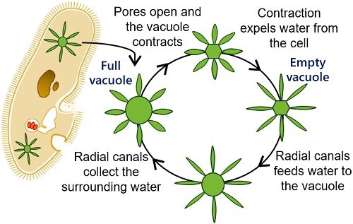
Around six to eleven radial canals are present around the contractile vacuole. These canals serve as “Feeding canals”. Thus, the radial canals feed all the wastes from the body into the contractile vacuole.
As a result, the contractile vacuole increases in size due to water accumulation. Contraction of vacuole results in discharging excess water through an anal pore. The anterior or posterior contractile vacuole works independently.