Inclusions in prokaryotes include certain types of food reserve granules and some monolayered non-unit membrane-enclosed cytoplasmic inclusions. Cell inclusion bodies can define as the pigmented molecules, which resides within the cell. It does not function like membrane-bound organelles but performs a pivotal role to store reserve materials.
Inclusion bodies occupy within the cytosol enveloped by a cytoplasmic membrane and an outer peptidoglycan cell wall. Prokaryotes possess a nuclear region, ribosomes and locomotory structures such as flagella, and pili that perform a distinct role within the cell.
Content: Inclusions in Prokaryotes
Definition of Cell Inclusion
Cytoplasmic inclusions can define as the non-living cytoplasmic aggregates, which are membrane-less and distributed throughout the cytoplasmic matrix. Unlike membrane-bound organelles, it does not participate in any metabolic reaction.
Inclusion bodies can define as the elementary bodies or cell remainings, which generally exist as storage granules, pigment molecules and secretory products. In prokaryotes, inclusion bodies categorize into three parts, namely gas vesicles, inorganic inclusions and food reserve bodies.
Types of Inclusion Bodies
The inclusion bodies in prokaryotes can be classified into the following classes:
- Gas vesicles: These are gas-filled structures used to regulate cell buoyancy of the bacteria living in lakes, oceans and seas.
- Inorganic inclusions: It includes metachromatin granules, sulphur globules, magnetosomes etc.
- Food reserve: In bacteria, food is reserved in the form of glycogen.
Gas Vacuoles
It is also called gas vesicles, which are generally found in cyanobacteria, purple and green bacteria and a few other planktonic forms. Gas vacuoles appear as the small, aggregated cylindrical structures. Its primary function is to protect the bacteria against potent radiations maintain cell buoyancy of the bacteria living in lakes, seas and oceans.
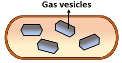
The gas vacuoles are nearly about 75 nm wide with pointed edges and approximately 200-1,000 nm long. Gas vacuoles characteristically occur in many cyanobacteria (Anabaena flos-aquae), few aquatic bacteria such as purple and green photosynthetic ones, and some archaebacteria such as Halobacterium and Thiothrix.
Bacteria possessing gas vacuoles can float at the depth to fulfil the requirements like appropriate light exposure, oxygen concentration, and nutrient uptake. But under high hydrostatic pressure, the gas vesicles collapse and then float upwards until the formation of new gas vesicles.
Gas vesicles resemble spindle-shaped and appear as a single membrane-bound gas-filled structure. The GvpA and GvpC proteins predominantly constitute the membrane formation by assembling themselves to form a layer of the gas vesicle. GvpA constitutes 97% to form a protein layer around the gas vacuole.
GvpC constitutes 3% of the protein-membrane and performs a key role in strengthening the wall of the gas vesicle. The membrane encloses the gas-filled hollow cylinder, which is hydrophobic but allows the transfer of atmospheric gases. The rigidity of the gas vesicle wall regulates the cell integrity against high pressure.
Polyphosphates
It is also called volutin or metachromatin granules. These structures retain the colour of methylene blue and appear reddish-purple. It occurs in the majority of bacteria and some microalgae, which accumulates inorganic phosphates in the form of polyphosphate granules.
Polyphosphate is an inorganic molecule that appears as a polymeric chain of orthophosphates joined via ester bonds. The existence of volutin granules was first studied in a bacterium (Spirillum volutans), which exhibits a metachromatic effect.
These granules comprise polymetaphosphates that helps in ATP synthesis and predominantly exists in Diphtheria bacillus and certain lactic acid bacteria.
Poly β-hydroxybutyrate (PHB)
It is one of the most common lipid inclusion bodies in bacteria, which forms by the joining of the β -hydroxybutyrate monomers via easter bond. Hence, poly β -hydroxybutyrate is a polymer of poly β -hydroxybutyric acid and appears as aggregated granules of width 0.2-0.7 µm.
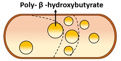
It forms by the condensation of acetyl CoA. PHB granules take up the colour of Sudan black. PHP lipid inclusions are visible under the electron microscope. PHB is commonly found in both archaebacteria and eubacteria.
It functions as a carbon storage reservoir, which provides long-term energy and participates in oxidative metabolism. Thus, the bacterial cells deprived of oxygen accumulate PHB granules to carry out fermentative metabolism.
Magnetosomes
It is one of the essential inorganic inclusion bodies, which appears as the polymeric chains of magnetite (Fe3O4). The bacteria that possess magnetosomes are considered as “Magnetotactic bacteria” which grows at very low O2 concentrations. Around 40-50 magnetite molecules combine to form a chain of magnetosomes, which have a diameter of 40-100nm. It forms intracellularly.
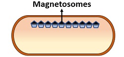
Some species from sulfidic habitats possess magnetosomes containing greigite (Fe3S4) and pyrite (FeS2). The shape of magnetosomes varies between species to species and may appear as square to spike-shaped.
Magnetosomes are encircled by a monolayer non-unit membrane. Generally, phospholipids, proteins, and glycoproteins constitute the membrane formation, in which proteins precipitate F3+ as Fe3O4 in the developing magnetosome.
Magnetosomes found in the bacteria found towards the sediment where O2 concentration is lower. Magnetotactic bacteria exhibit a property of magnetotaxis, by which they can move by the side of earth’s magnetic field, and also known as the living magnets.
Hence, each magnetosome will function as a tiny magnet to decide the southward and northward directions. Magnetosomes direct the movement of bacteria living in fresh and marine habitats so that they swim down to the optimum depth or nutrient-rich sediments.
Sulphur Globules
It includes photosynthetic purple sulphur bacteria and filamentous non-photosynthetic bacteria (Beggiatoa and Thiothrix) that grows in anaerobic and H2S rich environment. It is another inorganic inclusion body.
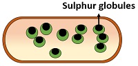
The bacterial cells oxidize H2S into elemental sulfur (H2S to S°), which in turn visible as the sulphur globules. Later, the stored sulfur in the granules oxidizes to sulphate (S° to SO42-) and the globules slowly disappear.
Sulfur globules exist within the periplasm instead of cell cytoplasm. The periplasm invaginates as the sulfur of the globules oxidizes and expands outwards to accommodate the globules.
Glycogen
Glycogen storage molecules are the reserve food material in prokaryotes. It is formed by the polymerization of glucose monomers, in which each unit is attached with the adjacent one via α(1-4) glycosidic bonds. Glycogen is the most common food reserve material are thoroughly scattered in the cytosol as 20-100nm wide small granules. It functions as a reservoir of carbon and energy.
Carboxysomes
These are polyhedral bodies commonly found in cyanobacteria, nitrifying bacteria and thermobacilli. It is also encircled by a 100 nm wide thin, non-unit membrane.
Carboxysomes contain ribulose-1, 5- bisphosphate carboxylase (RUBISCO) in a paracrystalline arrangement. It is believed that carboxysomes increase the production of RUBISCO, which in turn allow more CO2 fixation without altering the cell osmolarity.
The facultative autotrophic bacteria, which grow either as autotrophs or as heterotrophs cannot form carboxysomes. Thus, the carboxysomes give an idea of bacterial adaptation to the strict autotrophic environment.
Chlorobium Vesicles
It is also called chlorosomes, which predominantly found in obligate anaerobes Heliobacteria species. Chlorobium vesicles appear as flat cylindrical sheets associated with the plasma membrane. Because of its oval shape, they are also termed as ellipsoid vesicles. It lacks non-unit membrane and functions as a site of photosynthesis.
Conclusion
Therefore, inclusions are the cell deposits dispersed within the cytosol that perform a distinct role within the cell. Cytoplasmic inclusions also serve as a means of bacterial identification, which can be common in the majority of bacteria while few are restricted to particular species of bacteria.