The life cycle of Achyla species involves asexual or sexual modes of reproduction. The genus Achyla includes approximately 47 species and belongs to the group of oomycetes or water mould. Few species of Achyla are phytopathogens like Achlya conspicua and Achlya klebsiana.
Most of the Achyla species are aquatic, while a few grow on damp soil. Formation and detachment of gemmae from the vegetative body of Achyla species facilitates vegetative reproduction. The asexual reproduction in Achyla species commonly occurs via sporulation of motile zoospores.
Conversely, sexual reproduction in Achyla species is achieved occurs through the gametangial contact of antheridium and oogonium. This post describes the features of the genus Achyla and different growth phases (vegetative, asexual and sexual) in the life cycle of Achyla species.
Content: Life Cycle of Achyla
Features of Achyla
Before moving on to the life cycle of Achyla, let us discuss the structural features:
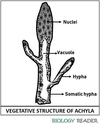
- Mycelium: Its mycelium appears as a white tangled mass. Each mycelium consists of branched hyphae that may emerge out in indiscriminate directions.
- Hyphae: The hyphae appear slender, broader at the base and gradually narrower at the tip. It may be coenocytic or aseptate. Hyphal projections help the Achyla species to penetrate the substratum.
- Nucleus: Achyla species possess numerous nuclei towards the edge of the cytoplasm surrounding the central vacuole.
Vegetative Reproduction
Members of the genus Achyla reproduce vegetatively by forming a dense or aggregated mass on the hyphae in the form of gemmae or thick-walled chlamydospores. The cytoplasmic mass of vegetative hypha transforms into a mass of spherical cells either at the terminal or intercalary position.
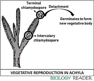
Chlamydospores are the resting spores densely filled with protoplasm, which develop on the coenocytic hyphae. In favourable conditions, the chlamydospores detach from the hyphal body through transverse septa. Later each chlamydospore may form a new vegetative structure.
Asexual Reproduction
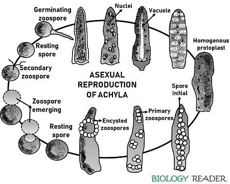
It occurs via sporulation of the motile zoospore that is summarized below. Zoospores appear pear-shaped with two apical flagella. They can also appear kidney-shaped with two flagella. A new vegetative body forms after the encystment and germination of zoospores.
Sporulation
Firstly, a sporangium originates after gradual swelling of the protoplasm towards the hyphal apex. Then, the sporangium initial being filled up with the multinucleate protoplasm. Later, the vacuole appears towards the centre, and the protoplasm propels toward the periphery.
Then, a sporangium separates from the hypha through a basal septum. During the first preliminary division stage, the central vacuole develops some ridge-like structures inside the dense protoplasm. The ridges are arranged in the form of intersecting lacunae, after which the sporangium divides into polygonal masses.
After that, a stage appears where the protoplasm achieves a homogeneous phase. The spore initials of polygonal masses are now ready to discharge out. Each spore initials are uninucleate. On maturity, spore-initials increase in size, become separated from each other and are termed primary zoospores.
Spore Encystment and Germination
The papilla of zoosporangium dissolves, and primary zoospores liberate one after one. Then, primary zoospores (non-motile) occupy the hollow sphere, where they encyst. Later, each encysted primary zoospore will break and develop a secondary zoospore after a few hours. Firstly, primary encysted zoospores remain in a resting phase for a while, and later they tend to emerge out.
Ultimately, the entire mass is discharged out to form a secondary zoospore. The secondary motile zoospore (biflagellate) swims for some time and again encyst except in Achyla aplanes. Finally, the zoospore will germinate in favourable conditions and give rise to the new hyphal system.
Sexual Reproduction
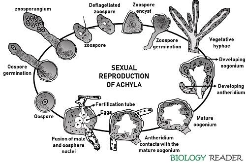
It is of oogamous type. In Achyla species, the male or female sex organs, i.e. antheridia and oogonia, are found on similar somatic hyphae. The majority of Achyla species are heterothallic, while few are homothallic. They undergo sexual reproduction through a method of gametangial contact.
Oogonium
Oogonia are the spherical structures that grow at the apical portion, either on the short lateral hypha or main hypha. Thus, the vegetative hypha that gives rise to the formation of oogonia is called a female or oogonial branch.
The female branch propels to develop a sac-like oogonial initial. The oogonial initials contain cytoplasm and nuclei in a homogeneous state, and later a central vacuole appears. After successive stages, the oogonium or swollen body separates out of the hypha by a basal septum.
Oogonia appear annular in shape and possess a thin, smooth and hyaline wall. Inside the oogonium, few nuclei degenerate, and the remaining divide mitotically to develop at least 2-6 oospheres. The mature oospheres appear spherical, uninucleate and devoid of a surrounding wall.
Antheridium
Antheridia develop on slender vegetative hyphae. They originate either from main hyphae or oogonial stalks. Antheridial protrusions grow in size, and the dense multinucleated protoplast shifts toward the swollen tip.
The swollen portion or antheridium separates via a transverse septum. The shape of antheridium is more or less club-shaped. Like oogonium, nuclei in the antheridium undergo mitotic divisions. Consequently, the antheridium interacts with the oogonial wall.
Fertilization
It occurs by the interaction of the mature antheridium and oogonium. An antheridium develops a fertilization tube that penetrates the oogonial cell and reaches an oosphere or egg. More than one antheridium can contact the oogonial wall via a fertilisation tube.
Then, karyogamy takes place by the fusion of the male nucleus with the nucleus of the oosphere. Fertilised eggs of the oogonium develop a thin wall.
Germination of Oospore
Oospores release out after dissolution of the oogonial wall. Then, oospores go into the resting phase for a while and germinate in favourable conditions. Upon germination, a thin layer surrounding the oospore bursts out.
After several meiotic cell divisions, the germ tube comes out of the inner protoplast into a branched and slender tube. After germination of oospore, the formation of zoosporangium produces zoospores, each of which germinates to develop new vegetative bodies.