Muscular tissue is a type of tissue present in the group metazoans, for the body movement or locomotion. Metazoans are the multicellular organisms belongs to the kingdom Animalia, whose cells differentiates to form tissues and organ. Muscular tissue provides mobility to the body organs of the organism.
Muscle tissues are of three types namely striated, unstriated and cardiac muscle which are structurally and functionally different from each other. Striated or skeletal tissues are attached to the bones by tendons and their contraction and relation are mediated by the central nervous system. Smooth muscles are present on the wall of hollow internal organs and their activity is controlled by the autonomous nervous system.
Cardiac muscle is located within the myocardium layer of the heart and its activity is controlled by the electrical impulses from the sinoatrial node or cardiac conduction system. The shape of skeletal, smooth and cardiac muscular tissues are characteristically different, as they possess cylindrical, spindle-shaped and branched fibre, respectively.
Therefore, the most common feature of muscular tissue is their ability to contractile through the actin and myosin proteins. Myogenesis is a process that occurs at the time of embryonic development which forms the muscle tissue. In this context, we will discuss the definition and different classes of muscular tissue along with the comparison chart of striated, unstriated and cardiac muscle tissue.
Content: Muscular Tissue
Definition of Muscular Tissue
The muscular tissue can define as the special types of white and red muscle tissues. The cells of the muscle tissue are called muscle cells. Muscular tissue originates from the embryonic mesoderm. It contributes almost half of the body weight. The cells of muscular tissue are long and narrow, due to which it is also termed as “Muscle fibres”. Its main function is to provide contractility or mobility.
Types of Muscular Tissue
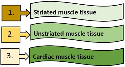
Muscular tissues are of three types:
- Striated muscle tissue
- Unstriated muscle tissue
- Cardiac muscle tissue
Striated Muscle
It is also known as voluntary muscle, as its functioning is under the control of a central and peripheral nervous system. Striated muscles contribute about 40% of the body weight. It attaches to the bones by tendons through both the ends. Due to the presence of many stripes (bands) in the striated muscles, they are also termed as “striped muscle”.
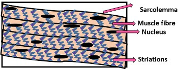
Structure
Striated muscle tissue has a complex structure that consists of many elements. These are found in the bundles that are made of few muscle fibres called “fasciculi”. All the fasiculies attach together by the connective tissue, and known as “perimycium”. The outer covering of the muscle bundle or fasciculi is made of connective tissues, known as “epimycium”.
- Size: Its length varies from 1-4 µm and diameter from 10-80 µm.
- Shape: Striated muscle is thread-like, elongated, cylindrical and unbranched.
- Elements: Its structure includes the following components:
- Sarcolemma: It is the transparent membrane surrounding muscle fibre. It has the inner plasma membrane of muscle fibres and a basement membrane of outer fibres.
- Sarcoplasm: The cytoplasm of muscle fibre is called sarcoplasm, inside which many flat and oval nuclei are present. Therefore, striated muscles are multinucleated or can be known as “syncytial”. There are some other structures also present inside the sarcoplasm like enzymes, sarcosomes (mitochondria), sarcoplasmal reticulum (endoplasmic reticulum), Golgi apparatus, glycogen and fat molecules etc. Myoalbumin, myogen and myoglobin are the three soluble proteins within the sarcoplasm.
- Sarcostyles: These are called myofibrils, which appears as the long contractile filaments. Its main function is muscle contraction and relaxation.
- Functions: Striated muscle helps in the body movement and also maintains the body posture.
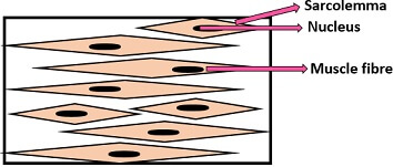
Unstriated Muscle
It is also called involuntary muscle, as its functioning is independent i.e. does not require the control of a central and peripheral nervous system. Unstriated muscles also interchangeable with the terms like smooth and visceral muscles because they are mainly found in the internal organs like alimentary canal, uterus etc.
Structure
Its structure is very simple. Unstriated muscles appear in the form of bundles and are made of muscle fibres. Inside the muscle fibres, there is a single, large, oval nucleus is present. Muscle fluid that surrounds the nucleus is called “sarcoplasm”. In the sarcoplasm, there are several filaments present that are arranged parallel and known as “myofibrils”. Actin and myosin filaments are the two common filaments of smooth muscle, which do not show dark and light band patterns. These filaments have an indefinite arrangement.
- Size: Length ranges from 50-200 µm and diameter from 2-5 µm.
- Shape: It appears as a long, thin, spindle-shaped and pointed at both the ends.
- Types: It is subdivided into two types:
- Helical smooth muscles: Here, the myofibrils are arranged helically and do not have a definite structure. Example: This type of muscle tissue present in the species of phylum Annelida and Mollusca.
- Paramyosin smooth muscles: Here, the actin, myosin and tropomyosin-B filaments are present in addition to myofibrils. Paramyosin occurs in the form of ribbons. It has a diameter of 15-150 µm. Example: This type of muscle tissue present in the species of phylum Mollusca.
- Function: Unstriated smooth muscle plays a key role in the propulsion of food through the alimentary canal and also helps in the contraction and relaxation of muscles.
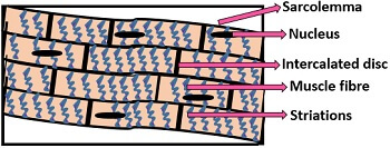
Cardiac Muscle
These are primarily found in the cardiac walls of the heart. Its structure is more or less similar to the striated muscle. But, its functioning is similar to the unstriated muscle that includes involuntary movements.
Structure
It is present in the bundles having branched muscle fibres that are surrounded by the sarcolemma. The muscle fibres bind to each other through septa and form a contractile network. In the cytoplasm, there are several integrated discs present in the irregular form. Numerous nuclei are also present inside the cytoplasm, which are flat and oval in shape.
- Size: Length ranges from 80-100 µm and diameter from 15-20 µm.
- Shape: These are long, thick, truncated and branched.
- Functions: Its main function is to pump blood throughout the body.
Comparison Chart
A table that is given below provides characteristic differences between the striated, unstriated and cardiac muscular tissues.
| Properties | Striated muscle | Unstriated muscle | Cardiac muscle |
|---|---|---|---|
| Also known as | Voluntary muscle | Involuntary muscle | Cardiac muscle |
| Location | Found attached to the bones | Found in the internal organs like stomach, liver, gall bladder, alimentary canal, urinary bladder etc | Found in the myocardium |
| Functioning | Performs voluntary movements | Performs involuntary movements | Performs involuntary movements |
| Control of muscle tissue | Controlled by central and peripheral nervous system | Controlled by autonomic nervous system | Controlled by sympathetic and para-sympathetic nervous system |
| Sarcolemma | Thick | Thin | Thin |
| Nucleus | Multinucleate | Uninucleate | Uni or Bi-nucleate |
| Presence of bands or striations | Present | Absent | Present |
| Branching | Branched | Unbranched | Branched |
| Intercalated disc | Absent | Absent | Present |
Muscle tissues are composed of many elements like protein, water, fats, carbohydrates, pigments, enzymes etc. The main characteristic feature of muscle is to convert the chemical energy into the mechanical energy from the beginning of muscle contraction to the end of muscle relaxation.