The difference between light and electron microscope is mainly due to the two properties like one is the source of illumination, and the second is the type of lens.
Source of illumination: It is the property of a microscope that ensures the clear visibility of the object or specimen and adds brightness to it.
Lens: It is used in the microscope, which can vary with different types of microscope available, and its primary function is to magnify the image.
The light microscope uses a direct source of light waves to form the image, whereas the electron microscope uses a beam of an accelerated electron. In the light microscope, the glass-based lens is used, and it has a combination of eye-piece, objective and condenser lens. In the electron microscope, the electromagnetic lens is used, and it has a combination of condenser, objective and projector lens. Here, we will discuss the key differences along with the comparison chart between the light and electron microscope.
Content: Light Vs Electron Microscope
Comparison Chart
| Properties | Light microscope | Electron microscope |
|---|---|---|
| Discovery | Given by Zoocharia Janseen in 1590 | Given by Ernst Ruska and Max Knoll in 1931 |
| Source of illumination | Light rays | Beam of electron |
| Lens used | Eye-piece, objective and condenser lens | Condenser, objective and projector lens |
| Lens type | Lens are of glass material | Lens are of electromagnetic material |
| Magnifying power | 1000X | 10,00,000X |
| Resolving power | 0.2 µm | 0.001 µm |
| Viewing screen | Image is viewed directly through eye-piece | Image is viewed on fluorescent screen |
| Power supply | Requires low power supply | Requires high power supply |
| Cooling system | Absent | Present |
| Sample preparation | Simple | Complex |
| Working | Easy to operate | Requires technical skills to operate |
| Types | Mainly of four types: Bright field, Dark field, Phase contrast and Fluorescence microscope | Mainly of three types: SEM, TEM and STEM |
| Vacumn system | Absent | Present |
| Cost | Cheap | Expensive |
| Magnification | Low, detailed structure cannot be studied | High, gives 3D structure of an object |
| Specimen used | 5 µm thick specimen can be easily visualized | Only thin specimen up to 0.1 µm can be visualized |
| Image obtained | Coloured | Black and white |
| Filament usage | Absent | Tungsten filament is used as an electron source |
| Radiation leakage | Absent | Present |
| For contrast of the image | Specimen is stained with dyes | Specimen is coated with heavy metals |
What is a Microscope?
A microscope is a device, which comprises a set of lenses that allow us to see the magnified view of an object or a specimen. This device helps us to study the internal and external structures of the specimen that would not be possible without the use of a microscope.
As from the name microscope, it is obvious that micro is a term used for minute things, and scope is a term used to look out things. Therefore, the microscope is an instrument that allows us to look or to see the microorganisms that are invisible to the naked eye. The study of the organisms under the microscope is called Microscopy.
Definition of Light Microscope
It is also called a compound microscope. A light microscope is an optical microscope, which uses a ray of light to view the image where a condenser collects the light and diverges it to the specimen. It has comparatively a low-resolution and magnification power than the electron microscope, which is about 0.2 µm and 500 – 1000 X, respectively.
Definition of Electron Microscope
It is an optical microscope, which uses a beam of an accelerated electron from the source of heated tungsten filament that transmits the electron to the specimen. It results in a high-resolution image of 0.001 µm resolving power, i.e. 250 times more than the light microscope and high magnifying power of 10,00,000 X.
Diagram of a Light Microscope
It consists of several components like:
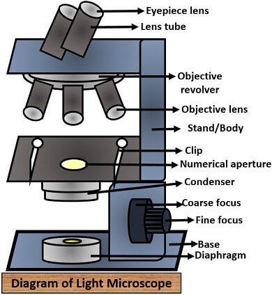
- Eyepiece lens: It is nearest to the eye of the observer. This lens is made of one or more lens. Observer observes the image that is first magnified by the objective lens, which is further magnified through the eyepiece. Its principal function is to convert the real intermediate enlarged image formed by the objective lens to the enlarged virtual image.
- Lens tube: It is the tube that holds an eyepiece and its length is about 160 mm but can vary with different types of microscope.
- Objective revolver: It holds multiple objective lenses with different magnifying power or capacity. One can spin or rotate this by one’s desire for the magnification of the specimen.
- Objective lens: This lens is nearest to the specimen or object, and it magnifies the image by collecting the light rays, then reflect it to the numerical aperture and finally gives a distinct view of the object.
- Clip: It holds the glass slide containing the specimen sample.
- Microscope stage: It provides a surface area, by which one can move the object slide to one’s desire or according to the part of the specimen one want to study or visualize.
- Condenser: It collects the light that incidents on it, which it projects back to the specimen for proper visibility.
- Fine and coarse focus: Both of these regulate the distance between the object and objective by moving the microscope stage. For the sharpness of the image, one can adjust both fine and coarse focus afterwards.
- Diaphragm: It adjusts the diameter of the light by preventing the image from overshine.
- Light source: For this, the light microscope uses light bulb-like LED.
- Stand or body: It holds all the components of the microscope.
- Base: It provides stability to the microscope.
Diagram of an Electron Microscope
It has several elements which are as follows:
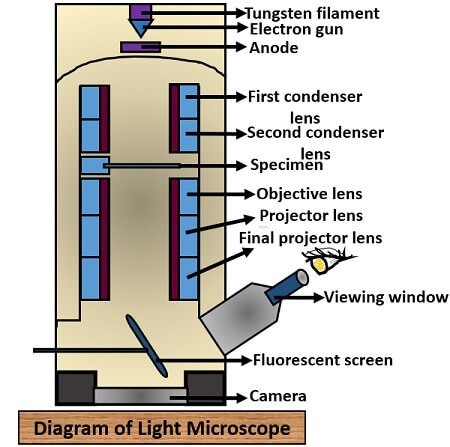
- Electron gun: It generates the beam of accelerated electron mainly through the tungsten filament by heating it to 100-1000 kV.
- Condenser lens: There is two magnetic condenser lens, which converges the light to the specimen.
- Objective lens: Magnetic objective lens focus the electron into an object and form the first real magnified intermediate image up to 2000 times.
- Projector lens: It further magnifies the real intermediate up to 240,000 or more times.
- Viewing screen: The electron microscope uses a zinc sulphate fluorescent screen or photographic plate to view the image.
- Camera: It is the charged coupled device that is located below the viewing screen.
- Specimen holder: Specimen is kept in a thin carbon film or collodion that holds by the metal grid.
Key Differences Between Light and Electron Microscope
- One of the characteristic difference is that a light microscope uses a light source, whereas an electron microscope uses a beam of an electron.
- The light microscope shows low magnifying and resolving power of 1000X and 0.2µm, respectively. In contrast, an e– microscope shows high magnifying and resolving power of 10, 00,000X and 0.001µm.
- The object is directly visible through the eyepiece in the light microscope. In contrast, the electron microscope makes the use of a fluorescent screen to see the enlarged view of the object.
- Electron microscope needs more power supply and technical skills to operate because of its complex construction, while a light microscope requires less power supply and its operation is easy.
- Vacuum system, tungsten filament, cooling system and radiation leakage is present in an electron microscope and absent in a light microscope.
- Sample preparation is easy in a light microscope in comparison to the electron microscope.
- Thick specimen up to 5 µm can be easily visualized in a light microscope, whereas only thin specimen up to 0.1 µm can be visualized in the electron microscope.
- The chemical dyes that are used for the staining of the specimens add contrast to the microscopic image and result in the coloured specimen. On the contrary, a specimen is coated with heavy metals that attract the beam of an electron, and it results in a black and white specimen in case of an e– microscope.
- A light microscope is broadly classified into a bright field, darkfield, phase contrast and fluorescent field microscope. An e– microscope is subdivided into TEM (Transmission Electron Microscope), SEM (Scanning Electron Microscope) and STEM (Scanning Transmission Electron Microscope) types of microscopes.
Conclusion
So finally, we have discussed the difference in properties, structure and components of both the light and electron microscope. Therefore, we can conclude that both the light and e– microscope works on the different principles, in which one uses a light source, and the other uses the electron to form the image. The construction and operation of both the microscopes are different, where a light microscope is easy to handle, and the electron microscope requires technical skills to operate.