Dimorphic fungi can define as a type of fungi, which has a dual life cycle. We can understand the meaning of the term dimorphic just by breaking it into two, in which Di means two and Morphic means morphology or structure. Therefore, dimorphic fungi possess two kinds of morphology in its life cycle. This dual character of dimorphic fungi is known as “Dimorphism”.
Based on the factors like temperature, pH, oxygen concentration, nutrients availability etc. the dimorphic fungi exists in two forms or structures, namely filamentous and yeast-like phase. Temperature is the primary factor of phase transition, and based on it, thermal and non-thermal are the two forms of dimorphic fungi.
The thermal and non-thermal dimorphic fungi infect humans, animals, rodents and insects and the latter are mainly phytopathogens. In this context, we will discuss the dimorphic behaviour and the two different phases of dimorphic fungi along with some important pathogens that are included in this group.
Content: Dimorphic Fungi
- Definition
- Dimorphic Life Cycle
- Phases of dimorphic fungi
- Examples of dimorphic fungi
- Transmission
- Diagnosis
- Treatment
Definition of Dimorphic Fungi
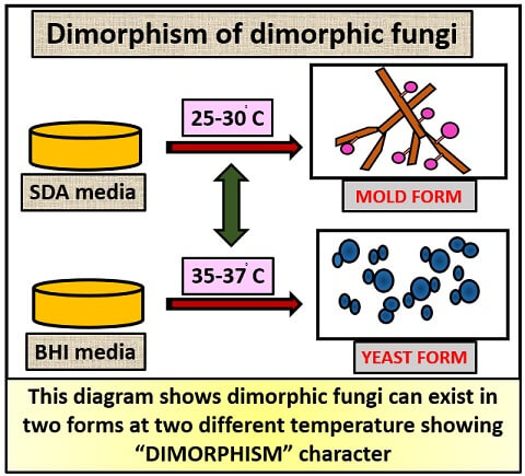
Dimorphic fungi possess a property of phase transition, in which they can reversibly change to the mould and yeast phase relative to the temperature. Mould phase occurs relatively at a low temperature (25-30 degrees C), whereas the yeast phase occurs remarkably at a high temperature (37 degrees C). As the temperature reaches 37 degrees C, the yeast phase transforms into a filamentous phase and vice versa. The species of dimorphic fungi belong to the phylum (Ascomycota), and all are thermally dimorphic except Coccidioides immitis.
Dimorphic Life Cycle
The dimorphic fungi exist in two forms at different environment, temperature, oxygen concentration etc. and the property exhibited by them is known as dimorphism. Filamentous and yeast-like phases are seen in their life cycle, which mainly depends upon the temperature and can be thermally dimorphic. Except for Coccidioides immitis, all are thermally dimorphic.
Phases of dimorphic fungi
- Mould phase
- Yeast phase
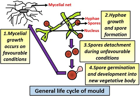
The general growth cycle of mould
It includes the following steps:
- First, a mycelial network forms.
- Then, the hyphae originate through the mycelial network.
- After that, hyphae form fruiting structures known as “Spores”.
- These spores then detach from the hyphae body during the unfavourable conditions.
- On the return of favourable conditions, the spore germinates and forms a new vegetative body.
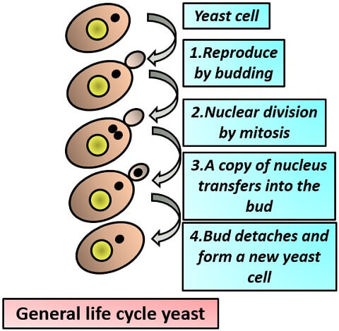
The general growth cycle of yeast
It includes the following steps:
- First yeast cell enlarges and forms a small protuberance, which extends outside the cell.
- This small outgrowth is called as “Bud”, and it forms by the phenomena called budding.
- After bud formation, the nuclear division occurs through mitosis.
- Then, a copy of genetic material passes to the bud.
- This bud then detaches during unfavourable conditions and forms a new yeast cell.
But in dimorphic fungi, these two growth cycle occurs one by one in its lifecycle:
In the mould phase :
- First, the spores detach from the vegetative cell during adverse conditions.
- Then, these spores remain in a dormant state on the soil.
- Then through wind or air, the spores go into the body and eventually, it turns into the yeast phase.
In the yeast phase:
- A fungal spore goes into the body or tissues of the host cell.
- This fungal spore then germinates into a yeast.
- Then, the yeast grows through budding and multiplies within the host cell.
- When yeast releases out of the host cell, it eventually returns to the mould phase.
Therefore, these two phases switch to one another, and the whole diagram is given below that is showing the life cycle of dimorphic fungi:
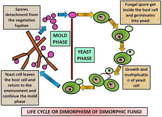
Table showing the growth conditions required in the mould and yeast phase:
| Phase of dimorphic fungi | Mold phase | Yeast phase |
|---|---|---|
| Conditions required for growth | Low temperature Optimal oxygen concentration Improved nutrients availability | High temperature Optimal oxygen concentration Improved nutrients availability |
| Nutrition type | Saprobic | Parasitic |
| Temperature range | <30degrees C | 35-40degrees C |
| Reproduction | Sporulation | Budding |
Examples of Dimorphic Fungi
Dimorphism of dimorphic fungi can be explained by taking a few examples like:
Histoplasma capsulatum
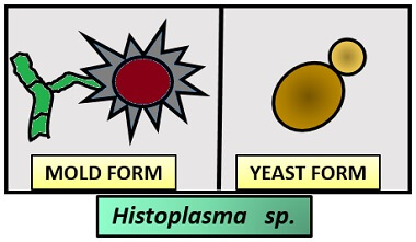
- Culture characteristics:
- In BHI media: Yeast phaseColony: Slow-growing
Size: Small
Shape: Oval
Colour: Cream coloured - In SDA media: Mould phase
Colony: Slow-growing
Appearance: Cottony
Colour: White to brown in colour
Hyphae: Thin, septate
Conidiophore: Small
Conidia: Single-celled, circular
- In BHI media: Yeast phaseColony: Slow-growing
- Distribution: Worldwide
- Habitat: It is found in the contaminated and nitrogenous soil enriched with bird droppings, chicken manure etc.
- Hosts: Human, donkeys, cats, dogs, horses etc.
- Target organ: Lungs, bones, skin, reticuloendothelial system etc.
- Disease: It causes histoplasmosis (an acute respiratory disease), which shows symptoms like illness, chest pain, cough etc.
Coccidioides immitis
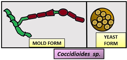
- Culture characteristics:
- In SDA media: Mould phase
Colony: Slow-growing, barrel-shaped
Appearance: Cottony
Colour: White
Hyphae: Filamentous and septate
Conidiophore: Small
Conidia: Also refers to Arthroconidia which are oval in shape - In BHI media: Yeast phase
Colony: Slow-growing, flat, and exist as Spherule.
Size: Small
Shape: Oval
Colour: Cream-coloured
Spores: Endospores present
- In SDA media: Mould phase
- Distribution: Cosmopolitan
- Habitat: Soil of low elevation desert
- Hosts: Human, cats, dogs, horses etc.
- Target organ: Skin and soft tissues
- Disease: It causes coccidioidomycosis (a pulmonary disease) that is also known as “Valley fever”.
Symptoms: Fever, chest pain, cough, sputum secretion, sore throat etc.
Blastomyces dermatitidis
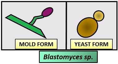
- Culture characteristics:
- In SDA media: Mould phase
Colony: Slow-growing
Appearance: Cottony
Colour: White to yellow in colour
Hyphae: Filamentous and septate
Conidiophore: Short and unbranched
Conidia: Single-celled, circular - In BHI media: Yeast phase
Colony: Slow-growing, broad base
Size: Small
Shape: Oval
Colour: Cream to tan-coloured
Cells appear as a fusiform budding form.
- In SDA media: Mould phase
- Distribution: Cosmopolitan
- Habitat: Soil with low pH (Acidic soil)
- Hosts: Human, cats, dogs etc.
- Target organ: Skin, bones, kidneys, CNS, eyes etc.
- Disease: It causes blastomycosis, which is also called North American Blastomycosis and sometimes Blastomycosis dermatitis.
Symptoms: Fever, chest pain, cough, excessive sweating, muscle and joint pain, lethargy etc.
Paracoccidioides brasiliensis
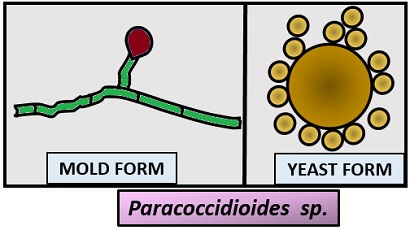
- Culture characteristics:
- In SDA media: Mould phase
Colony: Slow-growing
Appearance: Cottony
Colour: White
Hyphae: Thin, septate
Conidiophore: Small
Conidia: Single-celled, oval to circular in shape - In BHI media: Yeast phase
Colony: Slow-growing
Size: Large
Shape: Circular
Colour: Cream-coloured
Cells exist with multiple daughter buds.
- In SDA media: Mould phase
- Distribution: Cosmopolitan
- Habitat: Soil
- Hosts: Human, cats, dogs etc.
- Target organ: Skin, reticuloendothelial system, mucosal membrane etc.
- Disease: It causes paracoccidioidomycosis, which is also called South American Blastomycosis and sometimes Paracoccidioidomycosis granuloma.
Symptoms: Skin ulcers, localized lesions, abdominal pain etc.
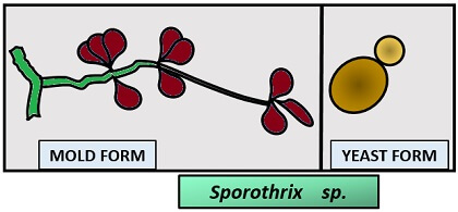
Sporothrix schenckii
- Culture characteristics:
- In SDA media: Mould phase
Colony: Fast-growing
Appearance: Velvety
Colour: Cream to orange grey in colour
Hyphae: Narrow and septate
Conidiophore: Slender and unbranched
Conidia: Rosette-like - In BHI media: Yeast phase
Colony: Slow-growing
Size: Small
Shape: Round to oval
Colour: Cream-coloured
Cells appear as a fusiform budding form.
- In SDA media: Mould phase
- Distribution: Worldwide
- Habitat: Soil with decomposed plants, woods, peat moss etc.
- Hosts: Human, cats, dogs, etc.
- Target organ: Subcutaneous nodules, lymphatics etc.
- Disease: It causes sporotrichosis or Rose handler’s disease.
Symptoms: Bumps (on fingers, hands, arms etc.), cough, chest pain, fever, headache, seizures etc.
Transmission
The transmission of diseases by dimorphic fungi can occur through the following ways:
- Inhalation of fungal spores through the air.
- By the direct contact with an infected person.
Diagnosis
Diseases caused by dimorphic fungi can be diagnosed by:
- Cell culturing methods.
- Serological testing like enzyme immunoassay, immunodiffusion test, complement fixation test etc.
- Chest X-ray.
Treatment
For the treatment of diseases caused by dimorphic fungi, some antifungal agents like Itraconazole, Fluconazole, Triazoles are being used in mild cases. In severe cases, Amphoterin-B is applicable.