Simple staining is one of the conventional staining techniques. From the name, it is quite clear that it is a very simple or direct staining method that uses a single stain only. The microorganisms are invisible to the naked eye, and to make them visible, staining is performed that gives divergence to a microscopic image. Direct staining makes the use of basic dyes like methylene blue, safranin, crystal violet, malachite green etc. called “simple or direct stains”.
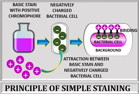
The basic stains possess a positive auxochrome that charges the stain’s chromogen particles to bind with the specimen. The chromophore group of the stain imparts colour to the microscopic image. As the basic stain carries a positive charge, it is also called positive or cationic stain. The purpose of direct staining is to add contrast to the specimen by directly stain the bacterial cells with a colourless background.
The simple stains make the organism visible and help us examine the organism’s shape, size, and arrangement, necessary to distinguish a particular group of organisms. The process generally involves three sequential steps like smear preparation, heat fixing and staining of the bacteria.
Content: Simple Staining
- Definition
- Simple Stains
- Principle
- Procedure of Simple Staining
- Video
- Advantages
- Disadvantages
- Conclusion
Definition of Simple Staining
Simple staining is defined as one of the ordinaries yet the popular method used to elucidate the bacterial size, shape and arrangement to differentiate the various bacteria groups. It stains the bacterial cell uniformly and thus increases the visibility of an organism. The term simple staining sometimes interchangeable with the terms like direct, positive or monochrome staining. Now let us understand why simple staining is called by such alternative names.
- Direct staining: Because it is a direct method that directly stains the bacterial cell with a colourless background.
- Positive staining: Because it uses positively charged basic dyes that bind with the negatively charged bacterial cell.
- Monochrome staining: Because it adds contrast to the specimen by using a single stain only.
Simple Stains
Simple stains can be defined as the basic dyes, which are the alcoholic or aqueous solutions (diluted up to 1-2%). These can easily release OH– and accepts H+ ion, due to which the simple stains are positively charged. As the simple stains are positively charged, they usually termed as positive or cationic dyes.
It is commonly used to colour most bacteria. As the simple stain carry a positive charge, it firmly adheres to a negative bacterial cell and makes the organism coloured by leaving a background colourless. Examples of simple stains include safranin, methylene blue, crystal violet etc.
The basic stains have different exposure times to penetrate and stain the bacterial cell.
| Basic stains | Exposure time to stain the bacteria |
|---|---|
| Methylene blue | 1-2 minutes |
| Crystal violet | 20-60 seconds |
| Carbol fuschin | 15-30 seconds |
| Safranin | 30-60 seconds |
Principle
Its principle is based on producing a marked contrast between the organism and its surroundings by using basic stain. A basic dye consists of a positive chromophore, which strongly attracts the negative cell components and charged molecules like nucleic acids and proteins. Thus, a simple staining technique results in a coloured bacterial cell against a colourless background.
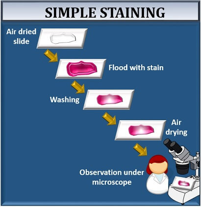
Procedure of Simple Staining
It involves the following three steps:
- Smear preparation
- Heat fixing
- Staining

Smear Preparation
A bacterial smear appears as a thin film of bacterial culture. For the smear preparation, we need to perform the following steps:
- Take a clean, grease-free glass slide.
- Add a drop of distilled water to the centre of the glass slide.
- Then, add inoculum from the bacterial culture with a sterilized inoculating loop on the glass slide.
- After that, mix the inoculum with a drop of distilled water to make a thin film by uniformly rotating the inoculating loop until a thin bacterial film is formed.

Heat Fixing
After smear preparation, move the prepared slides over the Bunsen burner’s flame at least three times. Then, allow the slide to air dry. There are many reasons to perform heat fixing, and it can not be skipped because:
- Heat fixing helps in the fixation of a specimen to the glass slide.
- Heat fixing helps the stain to penetrate the smear.

Staining of Bacteria
It is the last and the most crucial step, in which one can identify the morphological characteristics of the bacteria through microscopic examination, once the cells get stained. This stage involves the following steps, which are as follows:
- Add stain to the heat fixed smear.
- Allow the stain to stand for at least 1 minute so that it can penetrate between the cells.
- Wash off the glass slide carefully.
- Blot dry the slide with absorbent paper (do not wipe the slide).
- Examine the glass slide under the microscope from low to high power to get a magnified view of the specimen. One can also add a drop of oil immersion over the glass slide’s stained area to observe it under 100X objective.
Video
Advantages
- Simple staining is a very simple method to perform, which stains the organism by using a single reagent.
- It is a rapid method that reduces the performance time by taking only 3-5 minutes.
- Simple staining helps to examine or elucidate the bacterial shape, size and arrangement.
- It also helps us to differentiate the bacterial cells from the non-living structures.
- Simple staining can be useful in the preliminary study of the bacteria’s morphological characteristics.
Disadvantages
- It does not give much information about the cell apart from the bacteria’s morphological characteristics.
- Through simple staining, we cannot classify a particular type of organism.
Conclusion
Therefore, we can conclude that a simple staining method is the easiest way to colour the microscopic object as it uses a single basic stain. The results of simple staining are based on the type of basic stain that has been used.
The colour of a stain will decide the colour of a specimen that has to be identified. For example, when the bacteria retain the safranin colour, they appear pink-red, and the same goes with the other stains.
There is an attraction between the positive stain and the negative bacterial cell in simple staining, which results in the observation of coloured bacteria with a bright background.