The difference between axon and dendrite lies in their shape and branching. As well as, they differ in their length and function. Axons and dendrites are the cytoplasmic projections of the nerve cell body. Both play an essential role in the conduction of nerve impulses.
Shape: Axons maintain a constant radius. Whereas, dendrites are thicker near the cell body and taper towards the end.
Branching: Axons tend to branch distally or sometimes may develop side branches. In contrast, dendrites often branch at semi-regular intervals.
Length: Axons are long and their length may vary (one meter or more), while dendrites appear shorter in length.
Function: Axons perform a primary role in nerve impulse transmission. They pass on information from one neuron to different neurons, muscles and glands.
In contrast, dendrites receive the synaptic inputs from the axon terminal. Later, they propagate it throughout the cell body or soma.
This post describes the key differences between the axon and dendrite. Also, we will discuss the definition, comparison chart, structure and similarities between the two.
Content: Axon Vs Dendrite
Comparison Chart
| Properties | Axon | Dendrite |
|---|---|---|
| Alternative name | Nerve fibre | Dendron |
| Meaning | Axons are the cytoplasmic projections of the soma that appear as elongated fibres | Dendrites are the cytoplasmic projection of the soma that appear as short branches |
| Number | Neurons generally have one axon but may contain side branches or axon collaterals | Neurons possess many dendrites |
| Shape | It appears as an elongated fibre | It appears as a small branch |
| Length | Axons are relatively longer than the length of dendrites | Dendrites are short |
| Diameter | Uniform (width is identical throughout the length) | Variable (broad near the soma and taper at the end) |
| Origin | It originates from the conical axon hillock | It directly originates from the soma |
| Branching | Distally branched | Branched throughout |
| Surface | Smooth | Rough due to dendritic spines |
| Dendritic spines | Absent | Present |
| Myelin sheath | May present or absent | Absent |
| Nissl’s granules | Absent | Absent |
| Ribosomes | Absent | Absent |
| Neurofibrils | Absent | Absent |
| Terminal ends | Axon splits into fine branches (Telodendria) | Absent |
| Synaptic knob | Absent | Absent |
| Transport vesicles | Absent | Absent |
| Function | It transmits information from one neuron to the next via synapsis | It receives signals from the upstream neurons and passes to the cell body |
| Malformations in neuron’s cytoplasmic projections | Multiple sclerosis, traumatic brain and spinal cord injury, peripheral neuropathies etc. | Autism, depression, anxiety, down syndrome etc. |
Definition of Axon
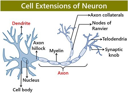
Axons or nerve fibres are long and slender, located between soma and axon terminals. Group A, B and C are the three types of axon. Group A and B are myelinated, and group C is nonmyelinated.
Axon originates from the initial segment or axon hillock that seems cone-like. The cytoplasm of the axon hillock comprises neurofilaments that cluster into fascicles.
Transmission of the nerve impulse from axon to axon terminals involves the following stages:
- The axon hillock serves as a site of initiation of an action potential. It integrates all the incoming signals from dendrites and soma.
- Potential travels down the axon. Note: Only if the strength of the impulse is greater than the threshold limit of the axon hillock.
- Later, the axon passes on this information from one neuron to the next.
Definition of Dendrite
Dendrites or dendrons are highly branched processes of the soma. These lack the myelin sheath. They are shorter in comparison to axons. In uni and bipolar neurons, dendrites resemble axons.
They appear broad near the cell body and taper when extended farther away from the cell body. Dendrite branching is a biological process wherein neurons form a dendritic tree. This facilitates the formation of new synapses.
Transmission of the nerve impulse from dendrites to soma includes:
- Dendrites function as the receiving units. At the synapse, they receive the released neurotransmitters (chemical signals) from the axon terminal.
- Later, these neurotransmitters bind to the dendrite’s specific receptors. Thus, dendrites provide a receptive surface for the synaptic inputs from the upstream neuron.
- Finally, they pass the synaptic inputs or nerve impulses to the soma.
Structure of Axon
Axolemma: It is a three-layered bi-lipid cell membrane that encircles the axon. Glia cells constitute the formation of the myelin sheath.
Myelin sheath: is a multilamellar spiral of a cell membrane. It is rich in fatty-acid substances. Axons may or may not have a myelin sheath.
Note: Myelin contains protein and fatty substances that usually form around the brain and spinal cord nerves.
Functions:
- Myelin sheath insulates the axolemma.
- It transmits electrical impulses all the way to axon terminals at a rapid rate. This is due to the nodes of Ranvier over the myelin sheath.
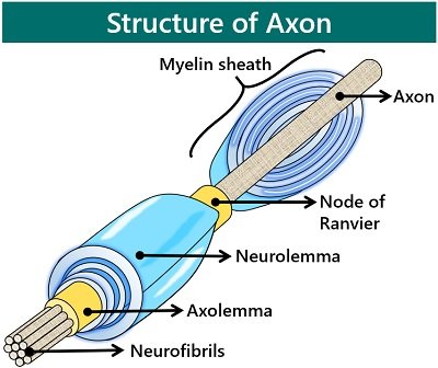
Axoplasm: It makes up the axon’s empty portion or cytoplasmic material. Axoplasm contains a cluster of microtubules and neurofilaments.
Within the cytoplasm, mitochondria are present in dispersed form. Beyond the initial segment, the axoplasm lacks RER and ribosomes.
Microfilament associated proteins (MAP) within the axons are of low molecular weight. Of all the types, tau is the most prominent MAP.
Axon collaterals: They are the side branches of axons. Axon collaterals resemble tree roots that split into terminal branches.
Telodendria: These are the axon’s terminal branches. Each telodendria has swollen ends that we call terminal or synaptic boutons. Axons connect with other cells at junctions known as synapses.
Structure of Dendrite
Receptors: The surface of dendrites contains an array of receptor proteins. These receive, process and transfer signals to soma. Receptors react to the neurotransmitters released from the upstream neurons.
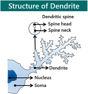
Dendritic spines: Dendrites resemble the main branch of a tree. They have several projections all over the surface that we call dendritic spines.
Dendritic spines appear as thorn-like protuberances. They function as the synaptic connections that contact with the nearby axons.
Dendritic spines enhance the surface area for synaptic contact. Microfilaments within the dendritic spines can modulate the spine’s shape.
Dendrite’s cytoplasm: They comprise several microtubules and fewer neurofilaments. The MAP like MAP2 has a high molecular weight and contain positive ends towards the cell body.
Mitochondria are longitudinally arranged in dendrites. Besides that, ribosomes and rough endoplasmic reticulum are also present.
Key Differences Between Axon and Dendrite
- Axons and dendrites are the cytoplasmic projections of the soma. The former appears as elongated fibres, whereas the latter appear like short branches.
- Neurons generally have one axon but may contain a few side branches or axon collaterals. However, a single nerve cell comprises many dendrites.
- Axons are relatively more extended than the length of dendrites. The diameter of an axon is uniform, i.e. width is the same throughout the length. In contrast, the diameter of dendrites is variable, i.e. broad near the soma and taper at the end.
- The axon’s surface is smooth. But, the surface of dendrites is rough due to dendritic spines.
- Dendrites have dendritic spines all over the surface, whereas axons lack such structures.
- Myelin sheath functions as an insulating layer. Axons may or may not have myelin sheath over their surface. On the other hand, dendrites lack this sheath.
- The axon’s cytoplasm contains several cytoskeletal elements, neurofibrils, transport vesicles, etc.
Unlike, the cytoplasm of dendrites includes microtubules, ribosomes, Nissl’s granules, SER, RER, etc. - Axon splits into fine terminal branches (Telodendria) with bulbous synaptic knobs. On the contrary, dendrites lack terminal branches.
- Axon transmits information from one neuron to the next via synapsis. But, dendrite receives signals from the upstream neurons and passes to the cell body.
Similarities
- Axons and dendrites are the components of a neuron.
- Both are the cytoplasmic projections of the soma.
- Both take part in the conduction of nerve impulses.
Conclusion
Neurons have one cell body and two projections (dendrites and axons). The neuron’s projections contribute to the nerve impulse conduction. Here, dendrites are receiving processes and an axon functions as a transmitting unit.
The direction of nerve impulse conduction is quite different. Axons work as signal transmitters, i.e. pass on the nerve impulse away from the nerve cell. While dendrites serve as signal receptors, i.e. bring nerve impulses towards the cell body.