The difference between rod and cone cells is primarily due to the difference in the factors like response to light stimulus, location within the retinal system of an eye, photopigments and the type of vision they form.
Response to light stimulus: Rods are more receptive towards light and function at scotopic (low) illumination levels. Conversely, cones are less receptive towards light and require high (day-light) illumination levels.
Location: Cones are highly concentrated in the fovea centralis, whereas rods are highly concentrated in the peripheral region of the retina.
Photopigments: Rods possess a single photopigment (rhodopsin), which breaks under the exposure of a wide bandwidth of light, i.e. rod photopigment is achromatic.
In contrast, cones have three distinct photopigments (L-opsins, M-opsins and S-opsins), which break down in a limited or specific bandwidth of light, i.e. cone photopigments are chromatic.
Vision: Rods form the scotopic or black and white vision, as they have colour insensitive and more light-sensitive rhodopsins. Oppositely, cones form photopic or coloured vision because they possess colour sensitive and less light-sensitive photopsins.
In this post, we will discuss the key differences between the rods cells along with the comparison chart. Also, the definition, structure and similarities between the two have been explained.
Content: Rod Vs Cone Cells
Comparison Chart
| Properties | Rod Cells | Cone Cells |
|---|---|---|
| Meaning | They are cylindrical photoreceptor cells lying outside the foveola of the retina | They are conical photoreceptor cells lying on the central region of the retina |
| Number in human eye | 120 million | 7 million |
| Location | Peripheral part of the retina | Fovea centralis of the retina |
| Size | Longer than the cones | Comparatively shorter than the rods |
| Type of vision | They are responsible for the scotopic, black and white or night vision | They are responsible for the photopic, coloured or day light vision |
| Light sensitivity | Sensitive to all wavelength of light (both direct and scattered) | Sensitive to a specific wavelength of red, green and blue light |
| Visual acuity | Low | High |
| Types | Only a single type of rods are found | Cones are classified typically into L, M and S cones |
| Photopigment or light-absorbing pigment | Rhodopsins | Photopsins |
| Outer segment | Outer segment appears long | Outer segment appears short |
| Membrane stacks in the outer segment | It contains more photopigment | It contains less photopigment |
| Inner segment | Narrow | Wider |
| Response time | Slow response and long integration time | Fast response and short integration time |
| Number of photons | Triggered by a single photon of light | Triggered by a large number of photons |
| Damage associated | Night blindness | Colour blindness |
| Function | They are important for retinal sensitivity | They are important for visual acuity (image details and colour specifications) |
Definition of Rod Cells
Rods refer to the light-sensitive photoreceptor cells in the retina, which are highly concentrated in the outer areas of an eye or outside the foveola, thereby responsible for peripheral or indirect vision. They function best in the low-light intensity and show more sensitivity towards light than the cone cells.
There are 120 million rods that appear cylindrical, elongated and a little leaner. Rods are responsible for the retinal sensitivity through which we can visualize things in dim light. Therefore, they give a scotopic or night vision under which everything seems in a grayscale range or black and white.
Definition of Cone Cells
Cones refer to the light-sensitive photoreceptor cells in the retina, localized in the central region (fovea centralis) of an eye, thereby responsible for the central vision. They function best in the high-light intensity and show less sensitivity towards light than the rod cells.
There are nearly 7 million cones that appear cone-like (shorter, broader and tapered). Cones are responsible for the visual acuity through which we can perceive colour and other object specifications. Thus, they give photopic or coloured vision.
Structure of Rods and Cones
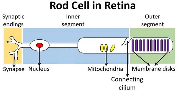
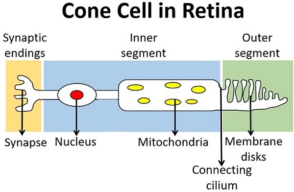
Both rods and cones possess four distinct structural elements.
Outer Segment
It holds the light-absorbing materials or photopigments that captures light or react to the stimulus of the photons. The photopigments transform the light stimuli into electrical impulses. Rods have more outer segment disks in the long outer segment and subsequently contain more photopigments.
Cones have less stacked membrane disks in the short outer segment, and subsequently contain fewer photopigments. The segment disks of rods contain the photopigments (rhodopsins), which absorb a wide range of light intensities.
The segment disks of cones differ, as the cones are of three types (L, M and S cones) that contain the photopsins pigments, which absorbs a spectrum of red, green and blue light, respectively.
The photopigments in the outer segment mainly perform phototransduction by firing an action potential althrough the neuron after receiving a light stimulus.
Connecting Cilium
It serves as a connecting link between the inner and the outer segment of the rods and cones. Connecting cilium is responsible for the disks morphogenesis in the photoreceptor’s outer segment.
Inner Segment
It encompasses the cell’s nucleus and organelles like mitochondria and Golgi bodies. Thus, the inner segment contributes to the cell body of the photoreceptors, and regulates the the membrane potential and energy metabolism. It receives the electrical signals from the photopigments and stimulates the synthesis of chemical signals or neurotransmitters.
Synaptic Terminal
It forms a synapse that connects the nerve endings to the axon terminal of the next neuron. The neurotransmitters from the inner segment reach the synaptic terminal of the rods and cones and eventually fuse with the plasma membrane and exit the cell.
Thus, the photoreceptors communicate with the axon terminal of the adjacent neurons (horizontal, bipolar and amacrine neurons) in the second or middle layer of the retina by specific cell receptors and neurotransmitters binding, and the process is called signal transduction.
The nerve impulse goes to the specialized ganglion cells (run along the retina’s surface) that form the optic nerves. Afterwards, the visual message goes to the other parts of the brain by the help of the glial cells (Muller cells, astroglia, and microglia) in the retina.
Key Differences Between the Rod and Cone Cells
- Rods are cylindrical-shaped photoreceptors lying on the peripheral retina, whereas cones are the conical-shaped photoreceptors lying on the fovea centralis of the retina.
- Rod cells are responsible for the scotopic and night vision, through which we could see objects in the grayscale range. Conversely, cone cells are responsible for photopic and day-light vision, through which we could perceive colour and finer details of the picture.
- Rod cells are sensitive to both scattered and direct light, whereas cone cells are only sensitive to direct light.
- Rods have no other types, whereas cones are of three distinct types, namely L, M and S, which absorbs red, green and blue light, respectively.
- A single photon can trigger the rod cells, while a large number of photons are necessary to trigger the cone cells.
- The visual impairments associated with the rods and cones are night and colour blindness. Rods possess rhodopsins that contains vitamin-A precursor whose deficiency causes night blindness, while cones comprise photopsins whose deficiency may consequence colour blindness.
Similarities
- Rods and cones are the modified nerve cells that are popularly known as photoreceptors.
- Both are localized in the outer photoreceptor layer of the retina.
- Both have photopigments or light-absorbing pigments in the form of transmembrane proteins, which appear as stacked membrane disks in the outer segment.
- Photons or light energy triggers the rods and cones cells.
Conclusion
Therefore, we can conclude that the rod and cone cells perform the same function. They both are the photoreceptor cells, whose photopigments capture or react to the light stimulus and trigger the phototransduction cascade.
As a result, the photopigments fire an action potential althrough the cell body or soma towards the synaptic terminal. Despite their function, the structure of rod and cone cells is similar, except for the overall shape.