Sclerenchyma tissue refers to one of the types of ground or simple permanent tissues, which possesses both primary and stiff secondary wall. They exist as rigid woody cell with a compact arrangement.
Sclerenchyma tissues aid cell integrity and conduction instead of being a dead cell. During the plant’s secondary, the sclerenchyma cells attain maturity and become dead cells due to lignin deposition.
Lignin restricts the exchange of water and gases, resulting in degeneration of inner protoplasm. This post discusses the definition, characteristics, types and functions of the sclerenchyma tissues.
Content: Sclerenchyma Tissue
Sclerenchyma Tissue Definition
Sclerenchyma tissue refers to the type of simple-permanent tissue, which initially exists as a living cell but becomes dead during the secondary wall development due to lignin accumulation.
“Lignification” refers to the phenomenon of lignin accumulation in the plant cells, which occurs after the completion of cell growth, and at the time of secondary thickening. Sclerenchymatous tissue predominates in the rigid areas of the plant body like leaf vein, stem, branches, trunk, bark etc.
Characteristics of Sclerenchyma Tissue
Sclerenchyma was derived from the Greek word “Scleros”, which means harder and “Enchyma”, which means infusion. A sclerenchyma tissue shows the following characteristic features.
- It is a dead, simple-permanent ground tissue.
- The function of sclerenchyma is similar to the collenchyma tissue, which aids mechanical support and tensile strength to the plants.
- Sclerenchyma cells function as a “Skeleton” of the plant system that contributes rigidity to withstand various ecological stresses.
- During the plant’s initial growth cycle, the sclerenchyma cells persist as the living cells and resemble spiral or ring patterns.
- During plant maturation, the sclerenchymatous cells become dead by the accumulation of lignin, making the cell harder and impervious to the exchange of water, solutes, gases, etc., between the environment and the inner protoplast.
- Sclerenchyma refers to a dead tissue because of its dead, degenerated or functionless inner protoplast.
- Mechanical and conductive sclerenchymatous tissues are two common types based on their function.
- Fibres, sclereids and tracheary elements are the three common types based on the morphology of sclerenchyma tissue.
- It comprises two layers of the cell wall; a primary cell wall and a thickened secondary cell wall (containing cellulose, hemicellulose, lignin etc.).
- The cell-wall type, rigidity, shape, size etc., of sclerenchyma will vary accordingly within different plant types.
Types of Sclerenchyma Tissue
Based on the function: A sclerenchyma tissue is broadly classified into two classes, namely mechanical and conductive sclerenchyma.
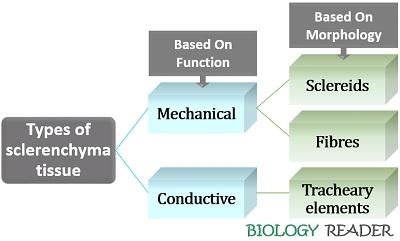
Mechanical Sclerenchyma
It is a kind of sclerenchymatous tissue that functions as a supportive tissue, which minimizes wilting in plants, maintains plant physiology, and provides strength to withstand the tearing forces of waves and current. Mechanical sclerenchyma comprises sclereids and fibre cells that contribute strength and stiffness to the plant system.
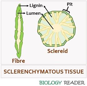
Sclereids
Sclereids refer to the mechanical tissues that occur singly or in groups. They are found associated with the plant’s vascular tissue, namely the xylem and phloem. Cell-wall thickening is non-uniform in sclereids.
They contain several simple pits with round apertures. Sclereids usually possess a narrow lumen. Based on the shape, the sclereid cells subdivides into the following classes:
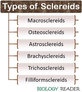
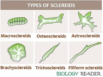
Macrosclereids
- They are also called “Malpigian cells”.
- Appearance: Elongated and columnar in shape.
- Cell wall: Thickened cell wall.
- Occurrence: Usually found in the outer epidermal cells of seed.
- Example: Seed coat of Pisum species.
Osteosclereids
- They are also known as the “Bone cells”.
- Appearance: It appears very similar to the shape of a bone of hourglass with enlarged, lobed and columnar cells. They are lobed towards the end.
- Cell wall: Thickened cell wall.
- Occurrence: Usually found below the epidermal layer, i.e. hypodermis of seeds and leaves of certain plants belonging to the Xerophytes category.
- Example: Leaves of Hakea species.
Astrosclereids
- They are also called “Stellate cells”.
- Appearance: They appear star-like and deeply lobed with the radiating arms from the central body. The radiating arms are usually pointed, irregular and varied in number.
- Cell wall: Thickened cell wall.
- Occurrence: Extends from the leaves’ upper epidermis to the lower epidermis.
- Example: Leaves of Thea, Olea etc.
Brachysclereids
- They are also called “Grit cells”.
- Appearance: They deeply resemble the parenchymatous cells and are roughly isodiametric.
- Cell wall: Thickened cell wall.
- Occurrence: It is commonly present in the fleshy portions of fruit.
- Example: Flesh of pear fruit, where brachysclereids form grit or stone cells.
Trichosclereids
- They are also termed the “Needle-like cells”.
- Appearance: They seem hair-like, more elongated, and branched cells stretching towards the intercellular spaces.
- Cell wall: Thickened cell wall.
- Occurrence: Present in the specialized tissues of leaves and roots.
- Example: Aerial roots of Monstera sp, leaves of olive and water-lily etc.
Filiformsclereids
- They are also called “Fibre-like cells”.
- Appearance: They are much elongated, sparingly-branched and uncommon kind of a cell.
- Cell wall: Thickened cell wall.
- Occurrence: Found in the specialized tissues of leaves.
- Example: Leaves of Olea.
Fibres
Fibres are other mechanical tissues that appear elongated and thick-walled with a narrow lumen and tapered ends. They generally constitute the plant’s ground and vascular tissues. Fibre sclerenchyma serves as a cell companion to the xylem and phloem tissues. They appear as independent strands or cylinders. Based on the shape, the fibre cells are categorized into the two following groups:
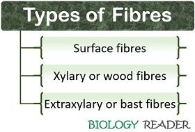
Xylary Fibres
They are associated with the primary and secondary xylem. The xylary fibres originate from the procambium, whereas xylary fibres originate from the cambium tissues of a plant cell. Xylary fibres exist in two common forms, namely libriform fibres and fibre tracheids.
- Libriform fibres have an elongated, thickened cell wall compared to the fibre tracheids and possess a simple pit with a longer pit canal.
- Fibre tracheids appear long, thick-walled and have a bordered pit with a smaller pit chamber.
Extraxylary Fibres
They are associated with the tissues outside the xylem (like phloem, cortex and pith of a plant cell). In monocots, the extraxylary fibre encircles the bundle sheath. They originate partly from the ground meristem and remaining from the procambium.
In dicots, the extraxylary fibres persist as independent bands or cylinders on the peripheral region of the vascular cylinder and innermost cortex layer. They originate wholly through the ground meristem tissues, whose structure, shape and composition are similar to the xylary fibres. The extraxylary fibres have three common types:
- Phloem fibres occur in the primary and secondary phloem of vascular plant tissues called “Bast fibres”.
- Cortical fibres are present in the cortex region of a plant cell that occurs singly or in groups and support the younger parts of plants.
- Perivascular fibres are present in the pericycle region of a plant cell, forming a vascular bundle cap of dicot and bundle sheath of monocots.
Conductive Sclerenchyma
It consists of a tracheary element, a peculiar property of vascular plants, which demarcates them from non-vascular plants. The tracheary elements provide both strength and water conduction.
Tracheary Elements
They occur in vascular plants and include vessel elements and tracheids.
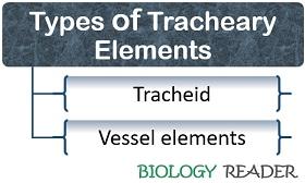
Vessel Elements
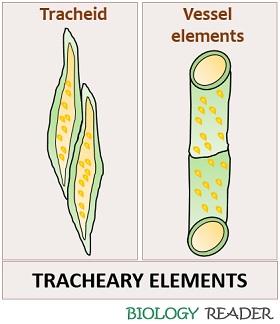
They possess perforated end walls (primary and secondary lignified walls) and present in primary and secondary xylem. Vessel elements are more efficient in water conduction, where the water flows vertically from one cell to the other without any hindrances.
They are a more specialized form of tracheary elements. Vessel elements have a small size than the tracheids. The vessel elements interconnect with the other vessels from one end of the cell to the cell’s other end in vertical rows.
Tracheids
They are the common cells in the xylem, which appear spindle-shaped and elongated with tapered ends. They participate in both water conduction and mechanical support. Tracheids appear elongated compared to the vessel elements and have a common feature in having a secondary wall thickening. They vary in shape (ranges from annular rings, reticulate etc., to pitted form).
Sclerenchyma Tissue Functions
Sclereids support the neighbouring tissues and protect the inner cells by forming a concentrating layer towards the periphery.
Fibre tissues contribute flexibility to plants. The septate fibres function as storage cells that reserve starch and oil droplets and protect the nearby inner tissues. The surface fibres facilitate seed and fruit dispersal. Plant fibres help in manufacturing textile, ropes, strings etc.
Tracheary elements or vessel elements solely participate in the water conduction. Tracheids have a high surface to volume ratio, protecting the plant from air embolisms or water stresses.
Good, it’s really helpful.