Adenovirus is a type of DNA virus, which appears medium-sized, non-enveloped and double-stranded. Adenovirus named after its isolation from the human adenoid tissue. It was first introduced by a scientist named Rowe, who cultured and isolated adenovirus via plasma tissue culturing. This group of virus can infect human, animals and birds.
Adenovirus can cause various diseases like respiratory tract infection, lung infections, common-cold, conjunctivitis etc. Adenovirus is ubiquitous, and it can disseminate worldwide. The life cycle of adenovirus is a complex process that begins with host-cell interaction, replication of viroid DNA, protein synthesis, bioassembly and host-cell lysis.
Adenovirus infections are most likely to occur in the late winter, spring and early summer months. It is most prevalent in children (between the age group of 6 months to 2 years) than the adults. In this context, we will discuss the definition, morphology, genome organization and classification of the adenovirus along with its epidemiology, pathogenicity and diagnosis. Besides, you will also get to know about adeno-associated viruses.
Content: Adenovirus
- Definition of Adenovirus
- Structure
- Genome Organisation
- Classification
- Epidemiology
- Pathogenicity
- Diagnosis
- Adeno-Associated Virus
Definition of Adenovirus
Adenoviruses belong to the group-1, ds-DNA human viruses, which usually cause acute respiratory infections and occasionally cause conjunctivitis, cystitis and gastroenteritis. It was first isolated after culturing the human adenoid tissue in the plasma tissue culture, after which it is named “Adenovirus”. It is a member of the family “Adenoviridae”, and it has two generas like:
- Mastadenovirus: It is the mammalian adenovirus, i.e. cause infections in mammals.
- Aviadenovirus: It is the avian adenovirus, i.e. cause infections in aves.
Resistance: Adenoviruses are relatively stable virus particles, which can survive at a temperature of 37 degrees Celsius for about a week. It gets inactivated at a temperature of 50 degrees Celsius or above. It also shows resistance towards chemical agents like ether and bile salts.
Host range: Adenoviruses can infect mammals and birds, and they are “Host-specific”. They can rapidly attack the human embryonic kidney, HeLa/HEp-2 cells.
Structure of Adenovirus
The size of adenovirus is larger than the other viruses, but its structure is quite simple, and it shows the following structural properties:
- Size: Adenoviruses are medium-sized.
- Shape: Adenoviruses show “icosahedral symmetry” and resemble the shape of the space vehicle.
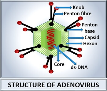
- Capsid: The adenovirus has an icosahedral, naked and non-enveloped capsid. The diameter of the capsid ranges from 90-100 nm. There are 252 protein subunits (capsomeres) in a capsid.
- Capsomeres: Adenoviruses comprise 240 hexon subunits and 12 penton subunits. The 240 hexon occupies the faces, and 12 pentons occupy the vertices of the capsid or shell.
- Pentons: Adenoviruses have a penton base and penton fibre. A long penton fibre attaches to the penton base and acts as hemagglutinin that carry specific antigens and is toxic to the cells. Knob like structures toward the terminal ends of the penton fibres promotes the virus’s attachment to the host cell.
- Core: Adenoviruses have a linear, ds-DNA genome. Protein V, VII and arginine-rich protein (µ) are the constituents that form the core of adenovirus. Inside the core of adenovirus, a protein of 55 KDa attaches to the 5’end of the ds-DNA.
Genome Organisation of Adenoviruses
They possess a linear, non-segmented and ds-DNA with a size of 30-38 Kb. The genome produces four types of gene products during gene expression.
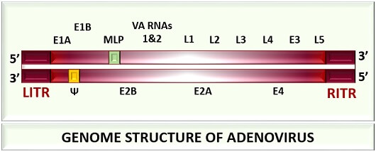
E1A is a gene product of the immediate-early phase in gene expression. The early-stage in gene expression produces gene products, namely E1B, E2A, E2B, E3 and E4. The intermediate stage of gene expression produces the intermediate gene product (VA) that encodes small RNAs (VA1 and VA2 RNAs). The late gene expression phase forms the late gene products (L1, L2, L3, L4 and L5, MLP and Ψ).
- Immediate early genes help in the replication of viral DNA.
- The early genes modulate the infected cells’ immune response and help in the viral DNA replication and transcription of late genes.
- An intermediate gene helps in regulating the mRNA translation.
- The late genes help in the synthesis of structural components at the time of virus biosynthesis and assembly.
The genome of adenovirus also encodes tripartite RNA leader or TPL sequences. Later, the TPL sequences are spliced up by the viral genome onto all the late viral mRNAs. Thus, both the DNA strands code specific genes that perform distinct tasks. Its genome also carries inverted terminal repeat sequences, due to which the denatured single strands transform into a circular or panhandle DNA molecule. To each 5’ end of viral DNA, a protein of molecular weight 55 KD is covalently attached.
| Phase of gene expression | Gene products of Adenovirus | Functional role |
|---|---|---|
| Immediate early phase | E1A | Binds with the human pRb and prevents from the cell cycle arrest |
| Early phase | E1B | Binds with the human polypeptide-53 and prevents apoptosis mediated by p-53 |
| E2A | Helps in adenovirus replication | |
| E2B | Helps in viral polymerization | |
| E3 | Removes CD95 from the from the cell surface and prevents TNF (Tumor Necrosis Factor) dependent apoptosis | |
| E4 | Helps in adenovirus replication | |
| Intermediate phase | VA (VA1 and VA2 RNAs) | Regulates the mRNA translation |
| Late phase | L3 | Cleaves viral precursor proteins |
| L4 | Helps in the viral replication | |
| L1 | Encodes capsid protein (p3a), which helps in the capsid biosynthesis | |
| L2 | Helps in DNA packaging and DNA condensation | |
| L5 | It is also a capsid protein encoding p-7 gene | |
| Ψ | It is a sequence giving signals during viral packaging | |
| Major late promoter (MLP) | Controls or promotes all the late phase genes | |
| - | LITR and RITR | Left inverted terminal repeat and Right inverted terminal repeat sequences contain origin of replication |
Classification
There are about 100 antigenic types of mammal and bird adenoviruses. From the 100 antigenic types, about 50 serotypes of adenovirus have been identified from human sources.
The human adenoviruses are categorized into six groups, depending on the properties like haemagglutination, fibre length, oncogenic potential etc. Let us study the six serotypes or subgenus of human adenovirus.
| Group (Subgenus) | Serotypes | Haemagglutination | Oncogenic potential (Tumourogenicity) |
|---|---|---|---|
| A | 12, 18, 31 | +/- (Partial haemagglutination) | High |
| B | 3, 7, 11, 14, 16, 21, 34, 35 | + (Complete haemagglutination) | Weak |
| C | 1, 2, 5, 6 | +/- (Partial haemagglutination) | - |
| D | 8, 10, 13, 15, 17, 19, 20, 22, 30, 32,33, 36, 39, 42, 47 | + (Complete haemagglutination) | - |
| E | 4 | +/- (Partial haemagglutination) | - |
| F | 40, 41 | +/- (Partial haemagglutination) | Not known |
Some of the human serotypes have been identified and isolated from the patients who concurred with AIDs. In adenovirus, group-specific antigens are present on the hexon, whereas type-specific antigens are present on the penton base and fibres. The immunological neutralization test can help us to study the serotypes of adenovirus.
Epidemiology
We can study adenovirus epidemiology by knowing all the factors related to the virus that can cause pathogenicity in the host cell.
- Reservoir or carrier: Humans and non-human primates (Monkeys) are the primary reservoirs.
- Transmission: Adenovirus may disseminate from person to person, through respiratory droplets, contaminated water and sometimes by sharing personal items.
- Clinical illness: It can cause fever (last for 3-5 days), atypical pneumonia, respiratory tract infection, tracheitis, bronchiolitis etc.
- Risk factors: Adenovirus mainly targets children and infants. Children are usually infected with mild pharyngitis or tracheitis. Infants are usually infected with fulminant bronchiolitis and pneumonia.
Pathogenicity
Adenovirus can cause the following pathogenic diseases:
Pharyngitis
It is a type of upper respiratory tract infection caused by viral serotypes 1-7. It is a clinical condition characterized by pharynx inflammation. Pharyngitis includes symptoms like:
- Sore throat
- Runny nose
- Fever
- Cough
- Headache
Pharyngitis symptoms can last for 3-5 days, and it includes complications like sinusitis and otitis.
Pneumonia
Serotypes 3 and 7 cause pneumonia in adults. Type 7 of adenovirus can cause serious illness in infants and young children. It is a clinical condition that causes inflammation in the lungs and also affects alveoli. Pneumonia includes symptoms like:
- Dry cough
- Chest pain
- Fever
- Difficulty in breathing
- Release of sputum
- Blue tinged eye
- Convulsions
Pneumonia can last for a few weeks to months and includes complications like asthma, diabetes, heart failure etc. It is also a type of upper respiratory tract infection.
Acute Respiratory Diseases
Mostly occur as outbreaks in military recruits and caused by serotypes 4, 7 and 21. It is a clinical condition that infects the nose, trachea and lungs and causes tracheitis, bronchiolitis etc. Acute respiratory infections obstruct normal breathing functions. It includes symptoms like:
- Congestion
- Runny nose
- Cough
- Sore throat
- Fatigue
- Dizziness
- Difficulty in breathing
Acute respiratory infections include complications like respiratory arrest, respiratory failure and congestive heart failure.
Pharyngoconjunctivital fever
Serotypes 3, 7 and 14 likely cause febrile pharyngitis and conjunctivitis in the civilian population. Pharyngoconjunctivital fever (PCF) is an acute and highly infectious viral disease of the conjunctiva, which leads to cause fever, pharyngitis and an acute follicular conjunctivitis.
Epidemic keratoconjunctivitis
Serotypes 8, 19 and 37 cause epidemic keratoconjunctivitis. It is a clinical condition that causes inflammation of the cornea and conjunctiva. Epidemic keratoconjunctivitis is highly contagious, which includes symptoms like:
- Swollen lymph nodes
- Fever
- Headache
- Fatigue
- Redness in the eyes
- Itchiness and irritation in the eyes
- Conjunctivital edema
- Excessive tearing
- Release of yellow discharge from the eyes
Acute follicular conjunctivitis
For this, serotypes 3, 4 and 11 are mainly responsible that cause inflammation of the conjunctiva, enlargement of submucous lymphoid follicles and preauricular lymph nodes.
Gastroenteritis
Serotypes 40 and 41 have been isolated from the faeces and designated as “Enteric type adenovirus”, which can cause infantile gastroenteritis. Gastroenteritis occasionally occurs and causes inflammation of the intestinal lining. It can be identified by the stool ELISA test.
Diagnosis
We can diagnose the pathogenicity of adenovirus commonly by the two methods:
- Tissue culture: First, collect the adenovirus from the throat, eye, urine, faeces etc., of an infected person. Then, inoculate the sample in the tissue culture and incubate it upto desired temperature and time period. The identification of an organism is possible by the following means:
- Observe the cytopathic effects
- Perform complement fixation test
- Perform neutralization test
- Electron microscopy also helps in the identification and diagnosis of enteric type adenoviruses. Immunoassay techniques like Fluorescent antibody test and ELISA also helps in the diagnosis. PCR based test also helps in the detection of viral DNA.
Adeno-Associated Virus
AAVs or Adeno-satellite viruses are other groups of a virus. They depend on the adenovirus for their replication and belongs to the family of Parpoviridae. AAVs are also classified as “Dependovirus”. Through electron microscopy, complement fixation or immunofluorescence methods, we can identify the AAVs. Its pathogenicity is still unknown.